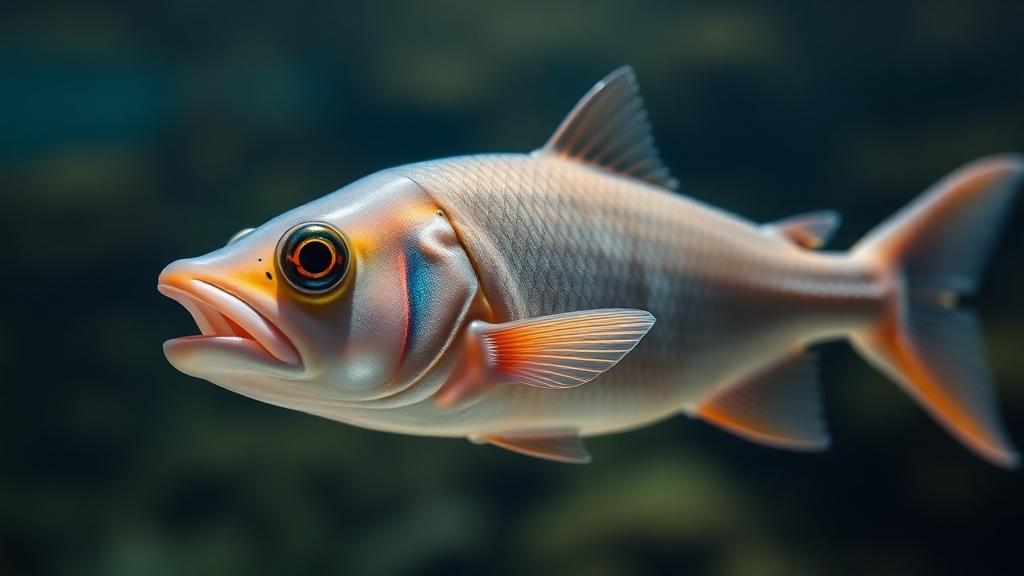
The red piranha is famous for its teeth, but a new study suggests its most intriguing biology may be happening inside a tiny organ buried deep in its brain. Researchers led by Keita Sato of Okayama University, together with colleagues at Kyoto University and Konan University, have discovered that the red piranha, Pygocentrus nattereri, carries two distinct copies of a light-sensitive protein called parietopsin, a pigment normally associated with the pineal gland, the small brain structure that lets many non-mammalian vertebrates detect light without using their eyes. The finding, published as an open-access paper in BMC Biology, offers a rare, functionally worked-out example of how the teleost whole-genome duplication, an ancient genetic event that reshaped the genomes of most modern fish, generated new molecular tools for sensing light. The work was published on 7 October 2026 and is among the first to show that duplicated opsin paralogs can diverge not just in the color of light they absorb, but in the very signaling messages they send inside cells.
Opsins are the protein workhorses of photoreception across the animal kingdom. Each opsin cradles a light-absorbing molecule derived from vitamin A, and the precise amino acid sequence of the protein tunes the wavelength of light that the complex absorbs most strongly. When a photon is captured, the opsin changes shape and triggers a cascade of chemical reactions inside the photoreceptor cell. Because opsins belong to the vast family of G-protein-coupled receptors, the downstream cascade matters enormously: some visual pigments activate cascades that break down cyclic AMP, a ubiquitous cellular messenger, while others raise it, and the choice of pathway fundamentally shapes how a cell responds to light. Understanding which pathway a given opsin uses is therefore essential to understanding what that opsin actually does for the animal, a point the new study places at the center of opsin evolution research.
The evolutionary backdrop is the teleost whole-genome duplication, often abbreviated TGD, a doubling of the entire genome that occurred in the ancestor of most living ray-finned fish. Genome duplications create spare copies of every gene, and those spare copies are free to mutate, acquire new functions, or fade away. For opsins, the TGD has clearly left its mark: duplicated paralogs of parapinopsin, vertebrate ancient opsin, and the long wavelength-sensitive opsin are known to differ from one another in absorption spectra, in where they are expressed, or in both. Yet, as the authors emphasize, the detailed evolutionary processes and mechanisms by which the TGD drove opsin diversification have remained poorly resolved. Most studies have focused on spectral tuning, the easiest property to measure, while the signaling behavior of duplicated opsins has been largely unexplored territory.
The team’s starting point was a survey of opsin genes across characins, the order of fish that includes piranhas, tetras, and their relatives. Several characins, they found, have retained two TGD-derived paralogs of parietopsin, which the researchers designated PT1 and PT2. This is unusual. Most teleosts, including the laboratory workhorse zebrafish, possess only PT1, while the Mexican tetra, Astyanax mexicanus, and various catfishes carry only PT2. The patchy distribution suggests that different fish lineages have kept different copies of this duplicated gene pair over tens of millions of years, a pattern that raises immediate questions about whether the two paralogs do different jobs and why some lineages abandoned one copy while others kept the other.
To answer those questions, the researchers characterized the two piranha proteins on three fronts: the wavelengths of light they absorb, the intracellular signaling pathways they engage, and the cells in which their genes are switched on. Measuring absorption spectra required expressing the proteins, reconstituting them with their light-sensitive chromophore, and spectroscopically probing the purified pigments. The results showed that the two paralogs are spectrally close but not identical. Red piranha PT1 absorbs maximally at 517 nanometers, in the green part of the spectrum, while red piranha PT2 peaks at 528 nanometers, a modest red-shift. Mexican tetra PT2 peaks at 517 nanometers, matching piranha PT1, and the PT2 of the Japanese catfish, Silurus asotus, absorbs at approximately 535 nanometers, the most red-shifted of the group.
The signaling experiments delivered the study’s most striking result. Using a heterologous cell-based assay, in which the fish opsins are expressed in cultured cells equipped with sensitive reporters, the team found that red piranha PT1 and PT2 couple to opposite signaling directions. When illuminated, PT1 increased the level of cyclic AMP in the cells, whereas PT2 decreased it. Neither parainopsin paralog, notably, produced a clear calcium response under the conditions tested. In practical terms, two proteins that descended from a single ancestral gene, absorb nearly the same green light, and sit in the same cells, push cellular chemistry in opposite directions when the lights come on. This kind of functional divergence, invisible to any study that measures only absorption spectra, is precisely what the authors argue has been missing from the picture of how genome duplication fueled opsin diversification.
The expression analysis added a further layer of surprise. Using fluorescence in situ hybridization, a technique that reveals exactly which cells contain specific messenger RNAs, the researchers showed that piranha PT1 and PT2 are co-expressed in the same pineal cells. Even more intriguingly, both are also co-expressed with parapinopsin 1, another pineal opsin whose duplicated teleost paralogs are known to have diversified. The pineal gland of the piranha therefore appears to be a densely multiplexed light-sensing tissue, packing at least three opsins, including two with opposing signaling polarities, into the same photoreceptive cells. How the cell integrates light signals that simultaneously raise and lower cyclic AMP is now an open and tantalizing question, one that could reshape assumptions about how simple, non-visual light detection works in fish.
The broader evolutionary implications reach well beyond piranhas. The retention patterns the team documented, with zebrafish keeping PT1, Mexican tetras and catfishes keeping PT2, and piranhas keeping both, illustrate that the fate of duplicated genes has played out differently across teleost lineages. Where both copies survive, functional divergence, in signaling if not in spectra, may be what allows them to coexist without redundant competition. Where only one copy survives, the lost paralog’s function may have been absorbed or rendered unnecessary. The study thus provides a concrete case study in a general principle: after genome duplication, the diversification of gene function can proceed through changes in signaling biochemistry, not merely through shifts in the physical properties of the protein’s light-sensitive pocket.
There is also a methodological lesson embedded in the findings. Had the researchers stopped at spectral measurements, PT1 and PT2 would have looked like near-identical twins, differing by a mere 11 nanometers in peak absorption. Only by assaying intracellular signaling did the deep functional split between the two paralogs come into view. The authors conclude that characterizing signaling properties is essential for understanding how the teleost genome duplication contributed to opsin diversification, and the implication for the field is clear: future surveys of duplicated opsins, in fish and beyond, will need to measure what these proteins do inside cells, not just what color of light they catch. As genome sequences accumulate from ever more fish species, studies of this kind promise to reveal how an ancient doubling of DNA, hundreds of millions of years ago, still shapes the way modern fish sense their world, from the green-lit shallows where tetras school to the murky rivers where piranhas swim with a light compass hidden in their brains.
Subject of Research: Functional diversification of duplicated parietopsin opsins after the teleost whole-genome duplication
Article Title: Two piranha parietopsins illuminate opsin diversification in teleosts
Article References: Sato, K., Yamashita, T., Sakai, K., Ohuchi, H., Kusakabe, T. G., & Gyoja, F. (2026). Two piranha parietopsins illuminate opsin diversification in teleosts. BMC Biology. https://doi.org/10.1186/s12915-026-02759-5
Image Credits: AI Generated
DOI: 10.1186/s12915-026-02759-5
Keywords: parietopsin, opsin, teleost whole-genome duplication, red piranha, pineal gland, photoreception, cyclic AMP, G-protein-coupled receptor, Mexican tetra, catfish, fluorescence in situ hybridization, BMC Biology
News Source: Juliet Wilcox. (October 7, 2026). Piranha Pineal Glands Reveal How Ancient Genome Duplication Diversified Fish Light Sensors. Scienmag.