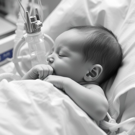
For premature and medically fragile newborns, removing a breathing tube is not a simple matter of switching off a machine. It is a high-stakes physiological test in which clinicians must determine whether an infant can sustain breathing independently without developing dangerous oxygen desaturation, carbon dioxide retention, apnea, or cardiovascular instability. A new study published in the Journal of Perinatology examines the biological signals that appear during this decision-making window in neonates older than 35 weeks’ postmenstrual age. Led by G.P. Lima, A. Kummaragunta, and J.G. Ortega, the research focuses on “physiologic signatures” observed during extubation readiness trials, or ERTs, offering a closer look at what an infant’s body may reveal before and during the transition away from invasive mechanical ventilation.
Mechanical ventilation can be lifesaving, but it also carries risks. An endotracheal tube bypasses the normal upper airway and connects the infant directly to a ventilator that delivers pressure and assists gas exchange. Prolonged ventilation may contribute to airway injury, inflammation, ventilator-associated infection, and damage to developing lungs. Removing the tube too early, however, can lead to respiratory failure and the need for reintubation, which is itself associated with additional stress and complications. This creates a narrow clinical balance: physicians must avoid both unnecessary prolongation of ventilation and premature extubation. ERTs are designed to help resolve that uncertainty by temporarily reducing or stopping ventilator assistance while clinicians monitor whether the infant maintains adequate respiratory function.
The study’s focus on neonates above 35 weeks’ postmenstrual age is clinically significant because maturity at birth does not necessarily predict readiness to breathe without support. Postmenstrual age combines gestational age at birth with the time elapsed since birth, providing a developmental measure that is particularly useful in neonatal medicine. Infants in this age range may include those born prematurely who have reached a more advanced developmental stage, as well as term or near-term newborns who remain ventilator-dependent because of respiratory disease, neurological impairment, infection, surgery, or other medical conditions. Their lungs, respiratory muscles, nervous systems, and cardiovascular responses may differ substantially, meaning that a single threshold—such as a particular oxygen requirement or ventilator setting—cannot fully capture readiness.
Physiologic signatures refer to coordinated patterns across measurable body functions rather than a single isolated number. During an ERT, clinicians may examine respiratory rate, tidal volume, breathing effort, oxygen saturation, heart rate, apnea frequency, blood gas measurements, and the infant’s response to changes in airway pressure. These variables interact dynamically. An infant can maintain a normal oxygen saturation for a period while using excessive respiratory effort, or show an acceptable breathing rate while failing to eliminate carbon dioxide effectively. Similarly, brief pauses in breathing may be harmless in one context but signal unstable respiratory control in another. By analyzing patterns over time, researchers hope to distinguish compensation from genuine physiological stability.
This approach reflects a broader shift in critical care medicine toward continuous, multidimensional monitoring. Traditional extubation decisions often rely on snapshots: a blood gas result, a ventilator setting, a chest examination, or the infant’s appearance at a particular moment. Yet breathing is a complex control system governed by the brainstem, respiratory muscles, lung mechanics, airway resistance, gas exchange, and cardiovascular circulation. A readiness trial challenges that system by removing part of the external support. The most informative signal may therefore be not a single measurement but the relationship between several measurements—for example, whether increasing respiratory effort is accompanied by falling oxygenation, rising carbon dioxide, or changes in heart-rate variability.
The work is also relevant to the growing use of electronic bedside data in neonatal intensive care units. Modern monitors record thousands of physiological observations, but the abundance of data does not automatically produce better decisions. Clinicians need validated patterns that can be interpreted reliably and connected to meaningful outcomes. Identifying reproducible signatures during ERTs could eventually support decision tools that alert staff to subtle deterioration before it becomes clinically obvious. Such systems would not replace neonatologists or respiratory therapists. Instead, they could help integrate streams of information that are difficult to assess simultaneously, particularly during busy periods when multiple infants require attention.
The importance of this research lies partly in its potential to improve the definition of extubation success. A trial may appear successful at the moment the tube is removed, yet an infant can deteriorate hours later as respiratory muscle fatigue develops or carbon dioxide gradually accumulates. Conversely, transient abnormalities may not always indicate that extubation will fail. A useful physiologic signature would ideally help distinguish these trajectories: stable adaptation to spontaneous breathing from temporary stress, and recoverable fluctuations from progressive respiratory collapse. That distinction could influence not only the timing of extubation but also the intensity and duration of monitoring afterward, including the choice of noninvasive support such as nasal continuous positive airway pressure or high-flow therapy.
The findings described by Lima and colleagues arrive at a time when neonatal care is increasingly concerned with individualized respiratory support. Two infants with similar gestational ages and similar ventilator settings may have very different reserves, muscle strength, airway mechanics, and neurological control of breathing. A physiology-based assessment could make extubation decisions more personalized by evaluating how each infant responds to reduced assistance rather than relying primarily on population-based thresholds. At the same time, the practical value of any proposed signature will depend on how consistently it can be measured across hospitals, devices, and clinical teams. Neonatal populations are heterogeneous, and factors such as sedation, recent procedures, infection, anemia, nutrition, and lung disease can alter physiological signals.
The study therefore contributes to an important clinical question rather than offering a universal formula for removing ventilatory support. Its central message is that readiness for extubation is a dynamic biological state, one that may be visible in the coordinated behavior of multiple signals before failure or success becomes unmistakable. Further research will be needed to determine which patterns are most predictive, how they perform in different neonatal populations, and whether incorporating them into bedside protocols improves outcomes such as reintubation rates, duration of ventilation, and length of intensive care. For now, the work highlights a promising direction in neonatal medicine: using the infant’s own physiology, captured continuously and interpreted in context, to make one of the most consequential transitions in intensive care safer and more precise.
Subject of Research: Physiologic signatures during extubation readiness trials in neonates older than 35 weeks’ postmenstrual age
Article Title: Physiologic signatures during extubation readiness trials in neonates >35 weeks’ postmenstrual age
Article References: Lima, G.P., Kummaragunta, A., Ortega, J.G. et al. “Physiologic signatures during extubation readiness trials in neonates >35 weeks’ postmenstrual age.” Journal of Perinatology (2026). https://doi.org/10.1038/s41372-026-02861-0
Image Credits: AI Generated
DOI: 10.1038/s41372-026-02861-0
Keywords: neonates, neonatal intensive care, extubation readiness trials, mechanical ventilation, respiratory physiology, premature infants, respiratory monitoring, extubation failure, postmenstrual age
Tags: apnea detection in premature newbornsbiological signals predicting successful extubationbiomarkers of breathing transition in preterm infantscardiovascular stability during neonatal weaning from ventilationneonatal extubation readiness assessmentneonatal oxygen desaturation during extubationneonatal respiratory stability markersneonatal ventilator weaning clinical decisionphysiologic signatures during neonatal extubationpostmenstrual age and extubation outcomesreintubation predictors in neonatal intensive carerisks of mechanical ventilation in neonates