In a retrospective proof-of-concept investigation published in CVIR Endovascular, vascular surgeons reported that physician-modified endografts constructed from an endoskeleton-based expanded polytetrafluoroethylene platform achieved complete technical success and sustained target vessel patency in high-risk patients presenting with complex aortic aneurysms. The clinical evaluation, conducted by Kenjiro Kaneko, Takao Ohki, and Eisaku Ito, examined four consecutive individuals presenting with complex aortic anatomy requiring a single visceral or renal branch fenestration. Every patient in the series presented with an infrarenal neck length of zero millimeters, rendering them entirely ineligible for conventional open surgical repair, standard endovascular aneurysm repair, or chimney graft configurations. Utilizing the commercially available AFX VELA platform, the surgical team performed back-table modifications to construct customized single fenestrations that were subsequently reinforced with expansile hydrogel coils. Over an average follow-up interval exceeding thirty-one months, the off-label endovascular approach maintained uninterrupted target vessel blood flow without precipitating procedural deaths, major adverse events, endoleaks, or secondary aortic reinterventions.
The management of complex aortic pathologies, specifically juxtarenal, pararenal, and thoracoabdominal aortic aneurysms, continues to present demanding clinical challenges due to the proximity of critical visceral and renal branch vessels. While fenestrated and branched endovascular repair has emerged as a predominant alternative, commercially produced custom-made devices require prolonged manufacturing periods spanning multiple weeks, which effectively precludes their timely deployment in urgent anatomical presentations. Standardized off-the-shelf branched endoprostheses eliminate manufacturing wait times but exhibit rigid anatomical instructions for use that disqualify more than half of prospective candidates, while simultaneously mandating full four-vessel branch reconstruction for localized pathologies. Under these restrictive circumstances, intraoperatively constructed physician-modified endografts offer an accessible therapeutic avenue that can be tailored directly to patient anatomy.
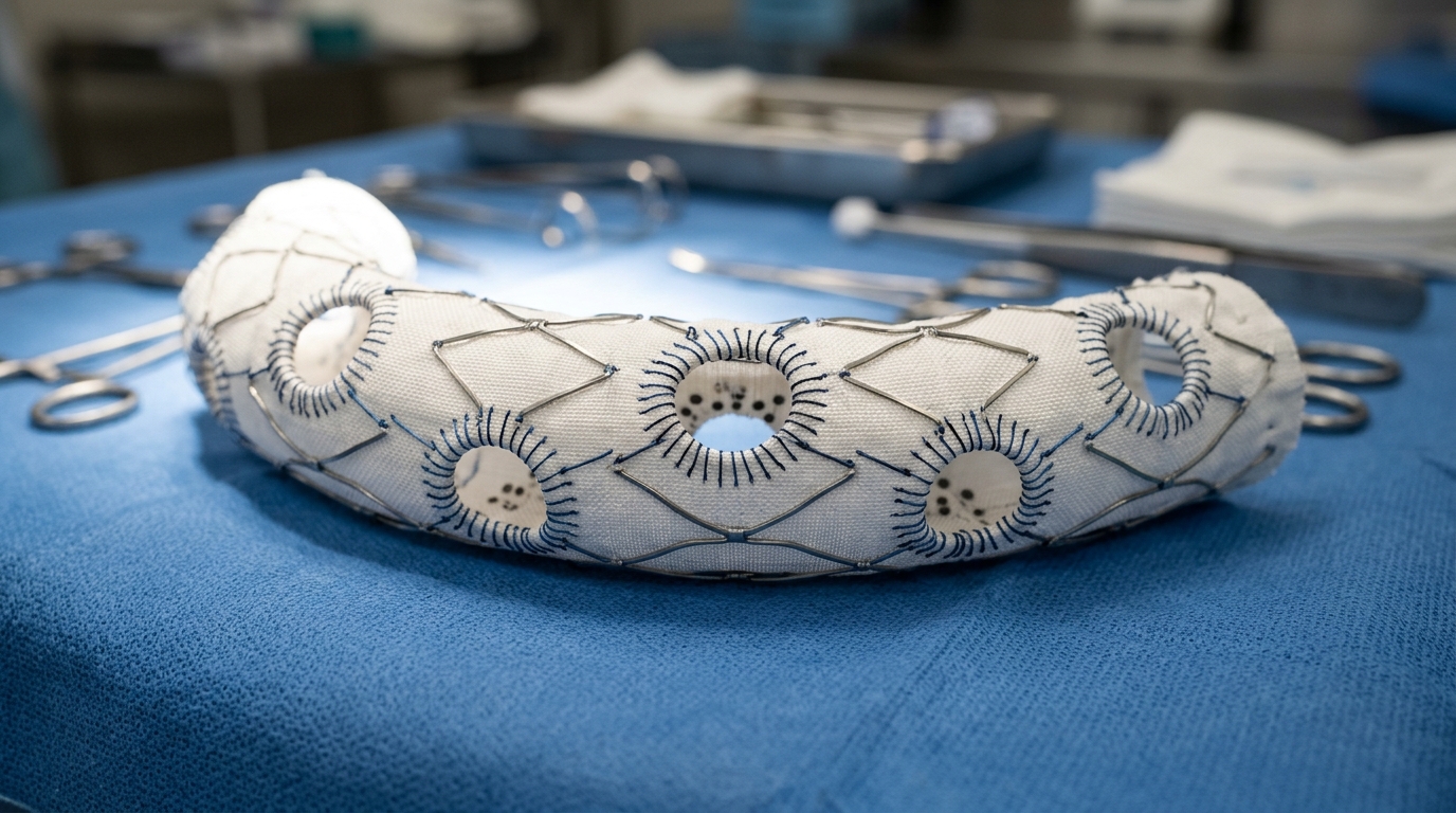
Despite the practical availability of physician-modified endografts, historical reliance on conventional polyester-based fabrics has introduced critical procedural hurdles during back-table preparation and systemic delivery. Traditional polyester endografts often present formidable resistance during manual resheathing into delivery sheaths, which can induce device infolding, compromised target vessel cannulation, and catastrophic procedural conversion to open laparotomy. Furthermore, mechanical fenestrations cut into woven polyester fabrics are prone to structural margin distortion, fabric recoil, and material degradation under repetitive physiological stress, raising persistent concerns regarding type IIIc endoleaks occurring at the junction between the fenestration margin and the bridging stent graft. To bypass these specific material vulnerabilities, the researchers explored the unique mechanical architecture of the AFX VELA platform. This device combines an internal metallic endoskeleton frame with an externally mounted expanded polytetrafluoroethylene fabric that is secured solely at its proximal and distal extremities, thereby allowing the fabric to be pulled free from underlying metallic struts during operative preparation.
The back-table modification protocol was executed under strict sterile operating conditions and required an average duration of 18.3 minutes, with procedural modification times ranging between 15 and 25 minutes across the four cases. The surgical team partially deployed the endograft by retracting the outer sheath up to the intended fenestration level, occasionally retracting the proximal release sleeve to obtain an unobstructed working space. Because expanded polytetrafluoroethylene fabric precludes thermal cauterization, a 7-0 polypropylene stay suture was applied to establish continuous fabric traction away from the internal metallic frame. Surgeons then performed sharp mechanical excision using a scalpel and Metzenbaum scissors to establish an orifice matched or sized one millimeter smaller than the native target vessel ostium. To preserve structural margin integrity, provide clear fluoroscopic visibility, and optimize bridging stent apposition, an expansile 0.018-inch AZUR peripheral hydrogel coil was circumferentially sutured along the fenestration margin using continuous expanded polytetrafluoroethylene sutures to prevent needle-hole seepage.
A central technical highlight of the modified endoskeleton platform was the simplicity of manual reloading back into the delivery system following fenestration creation and coil reinforcement. Because the metallic framework is situated on the inner aspect of the fabric, the external delivery sheath does not exert direct compressive friction against exposed metal struts, thereby permitting straightforward manual resheathing without the need for specialized mechanical crimping instruments or cumbersome auxiliary maneuvers. Although resheathing the segment housing the proximal release sleeve required operator familiarity, all four modified grafts were reloaded smoothly without evidence of fabric bunching or structural deformation. Immediately prior to vascular insertion, the integrity of each construct was confirmed under fluoroscopy, verifying that the sutured hydrogel coil had not undergone displacement or luminal distortion. This reproducible reloading capability directly addresses one of the primary rate-limiting technical hazards documented across conventional polyester-based physician-modified endograft series, where resheathing difficulties frequently precipitate intraoperative graft malfunction.
Intraoperative deployment of the modified endograft capitalized on the bidirectional expansion mechanics inherent to the VELA platform, providing surgeons with valuable flexibility regarding target vessel cannulation. Depending on patient-specific vascular anatomy, arterial access conditions, and branch take-off angles, the operative team selected either a distal-first retrograde approach or a proximal-first antegrade approach. For three patients exhibiting acute distal vessel angulation without severe thoracic mural thrombus, an antegrade strategy was implemented via right brachial artery access alongside bilateral femoral entries. Releasing the proximal front stop while maintaining constraint on the distal back stop secured an essential working channel between the aortic wall, the endograft, and the target branch orifice to facilitate wire and sheath passage. Conversely, in the single retrograde procedure selected for a cranially directed right renal artery, partial distal deployment under front-stop proximal constraint achieved an analogous working space, permitting smooth target cannulation prior to full proximal graft seating.
Detailed operative measurements recorded during the four interventions demonstrated a mean total operative time of 224.3 minutes, ranging from 203 to 243 minutes, with an average fluoroscopy exposure of 57.4 minutes. The procedures required a mean contrast medium volume of 343.0 milliliters and were associated with an estimated mean blood loss of 317.0 milliliters. The targeted visceral branches comprised two right renal arteries and two superior mesenteric arteries. Bridging stent reconstruction utilized a balloon-expandable GORE VIABAHN VBX endoprosthesis in three patients, flared within the aortic lumen using non-compliant balloons sized two millimeters larger than the target vessel diameter to prevent pull-out dislodgement. In the remaining patient, where a pararenal aneurysm sac left an anatomical gap to the right renal ostium, a flexible self-expanding VIABAHN stent was deployed to provide enhanced anatomical conformability. Adjunctive procedures included planned coil embolization of the celiac trunk in two patients after verifying mesenteric collateral flow through the gastroduodenal artery.
All four surgical procedures attained primary technical success, reflecting accurate device positioning, patent visceral branch reconstruction across the fenestrations, complete exclusion of the aneurysmal sacs, and the absence of type I or type III endoleaks on completion angiography. No patient required conversion to emergency open surgical intervention. During the initial thirty-day postoperative surveillance period, zero mortalities were recorded, and no patient suffered major adverse clinical events such as myocardial infarction, stroke, bowel ischemia, spinal cord injury resulting in paraplegia, or acute renal deterioration. Renal safety was demonstrated by the complete absence of a fifty percent reduction in baseline estimated glomerular filtration rate and the absence of newly initiated hemodialysis. The postoperative hospital stay was brief, ranging between four and six days with a mean duration of 5.3 days, and all four individuals were discharged home ambulating independently without requiring ongoing specialized institutional rehabilitation.
Over a mean clinical and radiological follow-up interval of 31.5 months, ranging from 17 to 49 months, the clinical benefits of the endoskeleton modification remained consistent and durable. Primary target vessel patency was maintained at 100 percent across all four reconstructed branches, with surveillance imaging confirming robust blood flow and no branch occlusions or stenosis-related interventions. Longitudinal follow-up computed tomography angiography and duplex ultrasonography detected no endoleaks of any category, confirming that no late type IIIc leaks emerged from the hydrogel-reinforced fenestration margins or modular connection sites. Aneurysm sac dimensions exhibited significant regression of at least five millimeters in two patients and remained completely stable in the other two, with zero instances of sac enlargement. Although one patient died from unrelated myelodysplastic syndrome at 41 months postoperatively, no aortic-related deaths occurred throughout the study, and stable postoperative renal function was maintained across all surviving patients.
While these preliminary outcomes demonstrate the technical feasibility and mid-term stability of physician-modified AFX VELA endografts, the authors emphasized several significant limitations inherent to their initial proof-of-concept experience. The study was restricted to a single medical center, lacked a comparative control cohort, and analyzed only four consecutive patients, precluding definitive statistical hypothesis testing. Additionally, combining an ePTFE endoskeleton platform with custom mechanical fenestrations and third-party bridging stents constitutes an off-label therapeutic approach whose long-term structural integrity remains unproven under continuous arterial pulsatile fatigue. The study intentionally included only saccular aneurysms requiring a single fenestration, leaving the technical feasibility of multiple fenestrations and extended fusiform aneurysm repairs unverified. The investigators cautioned that repetitive biomechanical loading could still induce late fenestration edge wear or component detachment over extended intervals, underscoring the absolute necessity for rigorous long-term patient surveillance and larger multi-institutional prospective trials to formally establish this endovascular technique.
Subject of Research: Physician-modified endoskeleton ePTFE endografts for complex aortic aneurysms
Article Title: Physician-modified endografts using an endoskeleton ePTFE platform for complex aortic aneurysms requiring a single fenestration: a proof-of-concept study
Article References: Kaneko, K., Ohki, T., & Ito, E. (2026). Physician-modified endografts using an endoskeleton ePTFE platform for complex aortic aneurysms requiring a single fenestration: a proof-of-concept study. CVIR Endovascular, 9(1), Article 117. https://doi.org/10.1186/s42155-026-00753-0
Image Credits: AI Generated
DOI: 10.1186/s42155-026-00753-0
Keywords: aortic aneurysm, endovascular repair, physician-modified endograft, ePTFE, fenestrated endovascular repair, vascular surgery, AFX VELA, target vessel patency, Physician-modified, endografts, endoskeleton, platform
News Source: Ophelia Keating. (November 6, 2026). Physician Modified Endoskeleton Endografts Show Early Success in Complex Aortic Aneurysm Cases. Scienmag.