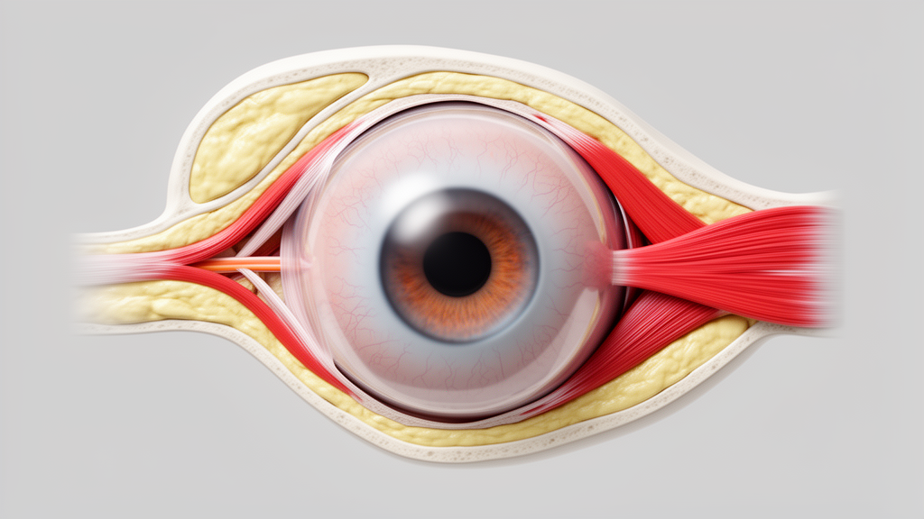
Researchers from Yokohama Brain and Spine Center and collaborating institutions reported that physical properties of the eye and surrounding tissues strongly influence directional differences in saccade motor characteristics. Published in Frontiers in Neurology, the study investigated why rapid eye movements vary in speed and timing depending on direction, a phenomenon previously observed but not fully explained. By measuring eye movements in 48 healthy participants, the team found that the speed of these movements is not uniform across all directions.
Instead, the data suggest that mechanical factors, such as elastic restoring forces and eyelid resistance, play a significant role alongside neural mechanisms in determining how the eye moves. The study focused on visually guided saccades, which are the rapid, voluntary shifts of gaze. Although saccades are generally believed to be generated by similar neural mechanisms in all directions, prior measurements have consistently shown directional differences in their movement characteristics.
The specific causes of these asymmetries remained unclear before this investigation. To address this, the researchers utilized an EyeSeeCam Sci 2 video-oculography system with a sampling rate of 500 Hz. This high-resolution setup allowed for precise capture of horizontal and vertical eye movements in a cohort of 28 males and 20 females, with a mean age of 40.38 years.
In the horizontal plane, the team measured saccades with amplitudes of 5, 10, 15, and 20 degrees in both centrifugal (away from the nose) and centripetal (toward the nose) directions. The results indicated that for larger amplitudes of 15 and 20 degrees, saccades in the medial hemifield were faster than those in the lateral hemifield. This pattern was observed in both centrifugal and centripetal directions.
Additionally, centripetal saccades were significantly faster than centrifugal ones. These findings suggest that the mechanical environment of the eye, rather than a simple neural command difference, contributes to the velocity profile of horizontal eye movements. Vertical saccades exhibited similar directional dependencies.
When measuring 5, 10, and 15-degree vertical movements, saccades in the upper hemifield were significantly faster than those in the lower hemifield for the largest amplitude of 15 degrees. This asymmetry was present in both upward and downward directions. Furthermore, centripetal saccades in the upper hemifield were faster than centrifugal ones, mirroring the horizontal findings.
The authors attribute these vertical differences to physical properties, including viscous resistance from eyelid coverage in the lower eye position, which may impede downward movements. Latency and amplitude analyses provided further insight into the mechanics of these movements. No clear trends were observed in horizontal saccade latency or amplitude across different directions.
However, vertical movements exhibited distinct patterns. Centripetal saccades showed shorter latencies, and upward saccades had larger amplitudes compared to downward movements. These subtle differences indicate that the initial phase of vertical eye movements is also influenced by the physical state of the periocular tissues.
The study notes that while neural mechanisms initiate the saccade, the final execution is modulated by the mechanical constraints of the eye socket. The authors specifically attribute the medial-lateral eye position difference to the interaction between saccades and vergence. The study emphasizes that saccade-vergence interaction is a key factor in explaining horizontal asymmetries.
Other directional asymmetries are attributed to physical properties such as the elastic restoring force in the centripetal direction. Additionally, viscous resistance from eyelid coverage in the lower eye position was identified as a factor slowing down downward saccades. The findings underscore the significant influence of periocular tissues on saccade motor characteristics.
While the brain sends neural commands to initiate saccades, the final execution of these movements is heavily modulated by the physical properties of the eye and its surroundings.
Subject of Research: Directional differences in saccade motor characteristics and the role of periocular tissue mechanics
Article Title: Directional differences in saccades
Article References: Jono, T., Takahashi, K., Hara, H., Kudo, Y., Schneider, E., Shiono, M., Tanaka, F., & Johkura, K. (2026). Directional differences in saccades. Frontiers in Neurology, 17, 1945647. https://doi.org/10.3389/fneur.2026.1945647
Image Credits: AI Generated
DOI: 10.3389/fneur.2026.1945647
Keywords: saccades, oculomotor control, eye movement, neuro-ophthalmology, periocular tissue, video-oculography, saccade velocity, horizontal saccades, vertical saccades, elastic restoring force, Directional, differences
News Source: Ophelia Keating. (October 23, 2026). Periocular Tissues Shape Saccade Speed Differences. Scienmag.