A new hypothesis suggests that intracranial aneurysm (IA) initiation is driven by pericyte dysfunction at the cerebral microcirculation barrier rather than by direct damage to the large-artery wall. Researchers propose that these cells, which maintain the blood–brain barrier, act as the earliest responders to abnormal hemodynamic stress. By integrating evidence from tumor biology, hypertensive small-vessel disease, and human genomics, the study outlines a trans-scale pathological sequence where pericyte-level disruptions bridge the gap between initial flow disturbance and subsequent structural failure in large intracranial arteries. This framework moves the focus from late-stage mechanical wall collapse to an earlier, potentially preventable window defined by cellular signaling defects.
Intracranial aneurysms, characterized by focal dilation of cerebral arteries, pose a high clinical risk with a case fatality rate near 40% following rupture. Current interventions are largely limited to surgical or endovascular procedures applied only after an aneurysm has already formed and become visible on conventional imaging. These macroscopic diagnostic methods lack the resolution to capture early molecular and cellular changes. The research suggests that by identifying these early cellular shifts, clinicians might one day intervene long before overt saccular bulging occurs, addressing an unmet clinical need in cerebrovascular management.
The proposed model describes a five-stage cascade starting with abnormal hemodynamics at circle-of-Willis bifurcations. Under physiological laminar shear, mechanosensitive channels known as Piezo1 and signaling pathways including TGFβ–ALK5 maintain pericytes in a quiescent, barrier-supporting state. However, in regions exposed to disturbed flow—characterized by low wall shear stress and high oscillatory shear—these mechanical signals are disrupted. Pericytes then undergo metabolic reprogramming, specifically an HK2-driven glycolytic shift that promotes pro-inflammatory conversion and increased contractility. This metabolic transition likely contributes to focal narrowing of the microvessels, which in turn feeds back to the bifurcation to establish a self-reinforcing loop of abnormal hemodynamics.
As pericyte function falters, the blood–brain barrier experiences dual mechanical and biochemical failures. Mechanically, pericyte detachment weakens essential N-cadherin contacts, while the loss of vitronectin signaling leads to endothelial junction disassembly. Biochemically, the damaged pericytes release inflammatory mediators such as IL-6 and MCP-1, which act on the adjacent endothelium. This stage of functional leakage occurs before the structural degradation of the large-artery wall, suggesting that cellular breakdown is an early, active contributor to the pathology rather than a late-stage consequence.
A critical component of this hypothesis is the spatial bridging between the microcirculation and the large-artery wall. The researchers propose that leaked plasma constituents and inflammatory mediators are conveyed through the perivascular space—a cerebrospinal fluid-filled channel—driven by pulsation-enhanced convection. This transport mechanism allows signals from the microcirculation to reach the adventitia of large intracranial arteries, where they encounter smooth muscle cells (SMCs). The study suggests that this external inflammatory load drives SMCs toward a dedifferentiated, synthetic phenotype and promotes the expansion of POSTN+ fibroblasts, leading to maladaptive fibrosis.
The structural remodeling observed in human IA tissue aligns with this outside-in signaling model. As SMCs convert to a synthetic, matrix-secreting phenotype, they contribute to disordered collagen deposition and passive wall stiffening, eventually exhausting the vessel’s elastic reserve. This is compounded by chronic cellular senescence, where both pericytes and SMCs acquire a senescence-associated secretory phenotype that releases matrix metalloproteinases, driving the destruction of the internal elastic lamina. This sequence highlights how fibrosis and IEL damage, once considered passive markers of aging or mechanical failure, may instead be early, active components of the aneurysm-forming process.
The authors emphasize that this model is a hypothesis-driven framework rather than established fact, with explicit grading of the evidence for each stage of the cascade. While the role of HK2 metabolic remodeling is well-documented in tumor vasculature and hypertensive models, direct demonstration in the context of cerebral pericytes under IA-relevant flow remains a priority for future study. Similarly, while the anatomy of the perivascular space and its potential for solute transport is supported by glymphatic research, direct spatiotemporal tracing in aneurysm models is necessary to confirm the bridging of signals from microvessels to the large artery.
Translational strategies suggested by the framework focus on stage-matched interventions aimed at delaying or slowing disease progression in high-risk individuals. Potential targets include the HK2 metabolic axis, the TGFβ/POSTN fibrosis axis, and the clearance of senescent mural cells via senolytics. The authors note that because these interventions act downstream of the initiating hemodynamic flow, their clinical value is limited to populations identified through computational fluid dynamics and emerging liquid-biopsy biomarkers. A successful preventative approach would require these molecular strategies to be carefully timed, as pathways like TGFβ are essential for baseline vascular homeostasis.
In conclusion, the study argues that reframing at least a subset of intracranial aneurysms as a trans-scale mural-cell disorder offers a clearer pathway for clinical validation. By identifying pericytes as the metabolic and signaling executors of hemodynamic stress, the model points toward specific experimental tests, such as in vivo tracing of leakage products and pericyte-restricted genetic modifications. The goal of this trans-scale approach is to shift the threshold for preventive care from the appearance of structural bulging to the detection of early, decompensated mural-cell function, potentially transforming aneurysm management from a surgical response to a proactive intervention.
The physical organization of cerebral blood vessels provides the architectural foundation for this microcirculatory concept. Intracranial arteries are suspended directly in the cerebrospinal fluid of the subarachnoid space and possess a thin adventitia lacking the perivascular adipose tissue of peripheral vessels. In the capillary network, functional integrity relies on an unusually dense mural population, where the ratio of pericytes to endothelial cells ranges from one to one up to one to three, far exceeding proportions in peripheral tissues. Despite sharing a common developmental origin from cephalic neural crest and cranial neuroectoderm, mature cerebrovascular mural cells occupy distinct anatomical compartments. At circle of Willis bifurcations, the vessel wall is dominated by medial smooth muscle cells, while capillary pericytes are restricted to neighboring leptomeningeal microvessels, distal perforating vascular beds, and sparse adventitial channels. This spatial segregation means abnormal mechanical forces must communicate across distinct microenvironments rather than through direct cellular migration.
Hemodynamic force transmission between these compartments is driven by sharp mechanical disparities. At arterial bifurcations, the apical carina endures laminar shear stress exceeding thirty dynes per square centimeter, whereas downstream lateral walls experience low oscillatory shear below five dynes per square centimeter. Aneurysms preferentially arise along these lateral walls, where pericytes face chronically unfavorable flow profiles. Although large intracranial arteries normally damp cardiac pulse waves, arterial stiffening and advancing age degrade this protective dampening. High-field magnetic resonance imaging demonstrates measurable pulsatility penetrating deep into lenticulostriate perforating arteries, with age-related losses in pulse attenuation. Computational fluid dynamics reveals that obtuse inlet angles between the middle cerebral artery and perforating branches aggravate local flow disturbances, funneling pulsatile energy into downstream pericyte-lined microvascular beds.
Once microcirculatory barrier breakdown occurs, transport physics across the perivascular space facilitates outward delivery of inflammatory mediators. Surrounding intracranial arteries, this fluid channel conducts net cerebrospinal fluid convection at velocities on the order of tens of micrometers per second. Because the distance between adjacent microvessels and large-artery adventitia spans several hundred micrometers to a few millimeters, convective delivery requires mere minutes. Small signaling proteins including interleukin-6 and monocyte chemoattractant protein-1 traverse these distances rapidly, with diffusion coefficients between ten to the negative sixth and ten to the negative seventh square centimeters per second. Larger macromolecules like albumin and fibrinogen are swept along by pulsation-driven solvent drag. Extravasated fibrinogen then functions as a damage-associated molecular pattern, activating Toll-like receptor four and Mac-1 integrins on resident macrophages. Because the cerebral arterial media lacks an extensive vasa vasorum and remains physiologically hypoperfused, minute amounts of delivered mediators achieve biologically potent concentrations.
This outside-in mechanism also addresses long-standing debates surrounding traditional pathogenic models. Classical pathology has long emphasized primary internal elastic lamina degeneration, attributing aneurysm formation to mechanical tensile exhaustion. The pericyte model accommodates this observation by positioning microcirculatory leakage as the upstream catalyst that floods the outer wall with matrix metalloproteinases and reactive oxygen species, accelerating elastic degradation. Similarly, while endothelial cells serve as initial mechanosensors through Piezo1 activation, the endothelial-pericyte unit functions as a coordinated circuit where pericytes amplify mechanical signals into metabolic and inflammatory shifts. Furthermore, high hemodynamic shear stress alone cannot fully explain aneurysm formation, as elevated shear is common across bifurcations that never develop lesions. Microvascular barrier breakdown offers the missing permissive trigger, depriving the overlying large-artery wall of vital microenvironmental homeostasis.
Validating this theoretical sequence requires targeted experimental and genetic investigations. Proposed experimental priorities include cranial-window two-photon in vivo imaging to trace fluorescent dextrans from leaky capillaries to the arterial adventitia in real time. Genetic manipulation using inducible driver lines, including Atp13a5 and platelet-derived growth factor receptor beta, could test whether pericyte-restricted hexokinase-2 knockout suppresses aneurysm formation under disturbed flow. Humanized microfluidic organoid models combining endothelial cells, pericytes, and smooth muscle cells under controlled shear can further bridge cross-species differences. These functional inquiries align with genome-wide association studies that identified seventeen intracranial aneurysm risk loci, showing marked variant enrichment in cerebrovascular pericytes and smooth muscle cells at significance levels exceeding ten to the negative fourth. Longitudinal clinical tracking combining flow imaging with circulating markers, including biotinidase and periostin, will ultimately test whether early cellular dysfunction reliably predicts future structural aneurysmal bulging.
Subject of Research: Pathogenesis of intracranial aneurysm initiation via pericyte-mediated microcirculation dysfunction.
Article Title: Pericyte dysfunction and perivascular-space signaling as triggers of intracranial aneurysm initiation: a hypothesis linking the cerebral microcirculation barrier to the large artery
Article References: Li, T., Zheng, J., Liu, W., & Shanshan, T. (2026). Pericyte dysfunction and perivascular-space signaling as triggers of intracranial aneurysm initiation: a hypothesis linking the cerebral microcirculation barrier to the large artery. Frontiers in Neuroscience, 20, Article 1978119. https://doi.org/10.3389/fnins.2026.1978119
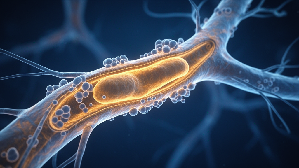
Image Credits: AI Generated
DOI: 10.3389/fnins.2026.1978119
Keywords: intracranial aneurysm, pericyte, microcirculation, hemodynamics, blood-brain barrier, fibrosis, perivascular space, mechanosensing, HK2 metabolic axis, cellular senescence, vascular remodeling, smooth muscle cell, glymphatic system, POSTN, computational fluid dynamics, cerebrovascular disease
News Source: Audrey Campbell. (November 18, 2026). Pericyte Dysfunction Linked to Intracranial Aneurysm Initiation in New Model. Scienmag.