


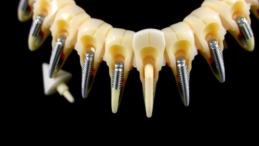
Biodegradable suture anchors made of PLGA are a mainstay of orthopedic soft-tissue fixation, but how their mechanical strength holds up as the material breaks down inside the body has been difficult to pin down. A new study from Zhejiang Pharmaceutical College and Ningbo Weierkaidi Medical Devices, published in Frontiers in Bioengineering and Biotechnology, maps that degradation-structure-mechanical relationship in detail and reports that a polycaprolactone (PCL) coating at a specific concentration can preserve compression performance through degradation-induced crystallization.
The researchers followed uncoated PLGA anchors through 24 weeks of in vitro degradation and observed a staged pattern. Mass loss was modest through the first eight weeks, staying below 5.4 percent, but accelerated after 16 weeks to more than 35 percent. Molecular weight told a different story: the number-average molecular weight (Mn) fell from roughly 185,000 Da to about 105,000 Da by 12 weeks and continued to decline thereafter, showing that chain scission was underway long before the bulk material visibly eroded.
That mismatch between molecular and macroscopic change is the study’s central finding. Compression Fmax, the maximum force the anchor could withstand, remained at or above its initial value through 16 weeks even as the polymer chains were steadily cleaving. The authors describe this as structural-mechanical decoupling, in which molecular degradation precedes any measurable macroscopic mechanical change rather than tracking it directly.
Imaging and thermal analysis added texture to the picture. Differential scanning calorimetry confirmed that the degrading material remained amorphous, with the glass transition temperature holding at approximately 65 °C throughout the degradation period. Scanning electron microscopy revealed progressive pore formation on the anchor surfaces, consistent with the staged mass loss profile.
The more applied part of the work tested whether PCL coatings at 5, 10, and 20 percent could regulate this mechanical evolution. The 10 percent coating performed best. It showed an initial melting enthalpy of about 46 J/g, corresponding to a crystallinity of roughly 33 percent, and that crystallinity increased further after four weeks. The authors interpret this as a crystalline physical crosslink network that preserves compression performance as the underlying PLGA degrades.
The coating concentration mattered in both directions. The 5 percent coating degraded too rapidly to provide lasting benefit, while the 20 percent coating showed no corresponding advantage over the 10 percent formulation. The result points to a narrow optimal window in which degradation-induced crystallization can be harnessed as a design principle for absorbable anchor coatings.
A preliminary in vivo assessment in a rat fascia-defect model, with only two animals per group at a Day 8 endpoint, examined biocompatibility. Blood and biochemical parameters showed individual deviations, and the implant caused notable local tissue reactions. Histology showed HE-stained inflammatory cell density lower in the sham group, at roughly 340 to 375 cells per square millimeter, than in the suture group, at about 743 to 813, and the anchor group, at about 775 to 882. CD206-positive densities remained below 10 cells per square millimeter in all groups. The authors note that these histological findings were descriptive, with no inferential statistics applied.
The study’s authors are explicit about its boundaries: the in vivo sample was small, the histology was descriptive, there were batch differences, and GPC molecular weight data came from single time points. The in vitro findings define the structure-mechanical relationship governing PLGA anchor degradation and support the PCL 10 percent coating as a design rationale, but the in vivo component is preliminary and does not establish clinical performance.
Subject of Research: Degradation behavior and mechanical preservation of PLGA suture anchors with PCL coatings
Article Title: Degradation-structure-mechanical relationships of PLGA suture anchors and PCL coating-mediated mechanical preservation via degradation-induced crystallization
Article References: Lu, Y., Xu, J., Li, N.-N., Hu, B., Xue, S., & Song, Q. (2026). Degradation-structure-mechanical relationships of PLGA suture anchors and PCL coating-mediated mechanical preservation via degradation-induced crystallization. Frontiers in Bioengineering and Biotechnology, 14, Article 2003985. https://doi.org/10.3389/fbioe.2026.2003985
Image Credits: AI Generated
DOI: 10.3389/fbioe.2026.2003985
Keywords: PLGA suture anchors, PCL coating, degradation-induced crystallization, biodegradable implants, orthopedic fixation, molecular weight decline, compression strength, crystallinity, biocompatibility, in vitro degradation, rat fascia-defect model, Frontiers in Bioengineering and Biotechnology
News Source: Neil Sanderson. (October 29, 2026). PCL Coating Keeps PLGA Suture Anchors Strong as They Degrade, Study Finds. Scienmag.



