Swiss scientists have used a cutting-edge method to stimulate neurons with light. They have successfully recorded synaptic transmission between neurons in a live animal for the first time.
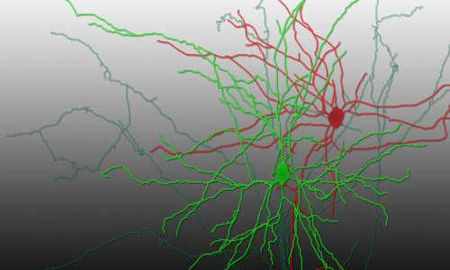
Reconstruction of a pair of synaptically connected neurons. Photo Credit: Aurélie Pala/EPFL
Neurons, the cells of the nervous system, communicate by transmitting chemical signals to each other through junctions called synapses. This “synaptic transmission” is critical for the brain and the spinal cord to quickly process the huge amount of incoming stimuli and generate outgoing signals. However, studying synaptic transmission in living animals is very difficult, and researchers have to use artificial conditions that don’t capture the real-life environment of neurons. Now, EPFL scientists have observed and measured synaptic transmission in a live animal for the first time, using a new approach that combines genetics with the physics of light. Their breakthrough work is published in Neuron.
Aurélie Pala and Carl Petersen at EPFL’s Brain Mind Institute used a novel technique, “optogenetics”, that has been making significant inroads in the field of neuroscience in the past ten years. This method uses light to precisely control the activity of specific neurons in living, even moving, animals in real time. Such precision is critical in being able to study the hundreds of different neuron types, and understand higher brain functions such as thought, behavior, language, memory – or even mental disorders.
Activating neurons with light
Optogenetics works by inserting the gene of a light-sensitive protein into live neurons, from a single cell to an entire family of them. The genetically modified neurons then produce the light-sensitive protein, which sits on their outside, the membrane. There, it acts as an electrical channel – something like a gate. When light is shone on the neuron, the channel opens up and allows electrical ions to flow into the cell; a bit like a battery being charged by a solar cell.
The addition of electrical ions changes the voltage balance of the neuron, and if the optogenetic stimulus is sufficiently strong it generates an explosive electrical signal in the neuron. And that is the impact of optogenetics: controlling neuronal activity by switching a light on and off.
Recording neuronal transmissions
Pala used optogenetics to stimulate single neurons of anesthetized mice and see if this approach could be used to record synaptic transmissions. The neurons she targeted were located in a part of the mouse’s brain called the barrel cortex, which processes sensory information from the mouse’s whiskers.
When Pala shone blue light on the neurons that contained the light-sensitive protein, the neurons activated and fired signals. At the same time, she measured electrical signals in neighboring neurons using microelectrodes that can record small voltage changes across a neuron’s membrane.
Using these approaches, the researchers looked at how the light-sensitive neurons connected to some of their neighbors: small, connector neurons called “interneurons”. In the brain, interneurons are usually inhibitory: when they receive a signal, they make the next neuron down the line less likely to continue the transmission.
The researchers recorded and analyzed synaptic transmissions from light-sensitive neurons to interneurons. In addition, they used an advanced imaging technique (two-photon microscopy) that allowed them to look deep into the brain of the live mouse and identify the type of each interneuron they were studying. The data showed that the neuronal transmissions from the light-sensitive neurons differed depending on the type of interneuron on the receiving end.
“This is a proof-of-concept study,” says Aurélie Pala, who received her PhD for this work. “Nonetheless, we think that we can use optogenetics to put together a larger picture of connectivity between other types of neurons in other areas of the brain.”
The scientists are now aiming to explore other neuronal connections in the mouse barrel cortex. They also want to try this technique on awake mice, to see how switching neuronal activity on and off with a light can affect higher brain functions.
Story Source:
The above story is based on materials provided by Ecole Polytechnique Federale de Lausanne.

