The finding, published as a case report in the journal General Thoracic and Cardiovascular Surgery Cases, came from frequency-domain optical coherence tomography performed two weeks after coronary artery bypass grafting in a 75-year-old woman. The structures appeared as signal-poor tubular channels within the adventitial layer of the graft, and three-dimensional reconstruction showed that they ran continuously along the vessel wall. The authors are careful to frame the observation as hypothesis-generating rather than definitive: the imaging could not confirm blood flow, functional patency, or the histological identity of the structures, and no baseline imaging existed to prove that pre-existing vasa vasorum had been preserved rather than newly formed or remnant.
The no-touch saphenous vein graft technique, in which the vein is removed with a thick pedicle of surrounding tissue, has demonstrated superior long-term patency compared with conventional harvesting in previous work cited by the authors. The proposed mechanisms include preservation of perivascular adipose tissue and adventitial integrity, which may help maintain the vasa vasorum, as well as avoidance of high-pressure distension, which may reduce endothelial and intimal injury. Because the vasa vasorum network is primarily located within the adventitia and adjacent perivascular tissue, the authors reasoned that careful protection of the adventitia alone might retain elements of this microvascular architecture even without a bulky tissue pedicle.
That question matters because the no-touch approach has a practical drawback. Harvesting the vein with a thick pedicle of surrounding tissue may increase leg wound complications and is not universally adopted, the authors note in their background section. Whether adventitial structures consistent with vasa vasorum remain detectable in carefully harvested conventional grafts without a thick tissue pedicle had remained uncertain, and that uncertainty is precisely what this single case was designed to probe.
The patient in the report had severe aortic stenosis and multivessel coronary artery disease and underwent aortic valve replacement together with coronary artery bypass grafting. The saphenous vein was harvested from her right lower leg using Metzenbaum scissors and an electrosurgical unit operated in DRY CUT and FORCED COAG modes, both set at 30 watts. Dissection was performed under direct visualization of the external surface of the vein wall, with an approximately 1 to 2 millimeter rim of surrounding adipose tissue retained to avoid injury to the adventitial layer. Side branches were ligated and divided approximately 1 to 2 millimeters from the graft trunk to minimize injury to the adventitia and its associated microvascular structures.
Just as important as the dissection was what the surgeons did not do. After harvest, the graft was gently dilated using arterial-line pressure from the cardiopulmonary bypass circuit, approximately 100 millimeters of mercury, rather than manual syringe distension. High-pressure manual distension has been associated with endothelial and intimal injury in the literature the authors cite, so the dilation strategy was chosen to keep mechanical stress on the graft wall as close to physiological as possible. The graft was then used for coronary bypass grafting in the standard fashion, and the harvesting procedure itself was recorded in a supplementary video file accompanying the article.
Two weeks after surgery, routine postoperative coronary angiography was performed, and frequency-domain optical coherence tomography imaging was obtained using a Dragonfly OpStar imaging catheter and SJM FD-OCT Imaging System from Abbott Cardiovascular. The imaging covered an approximately 60-millimeter proximal segment of the graft, and within that segment an approximately 10-millimeter region containing structures consistent with vasa vasorum was selected for three-dimensional reconstruction. Structures consistent with vasa vasorum were defined as signal-poor tubuloluminal structures identified on serial cross-sectional and longitudinal FD-OCT images and located within the adventitial or immediately perivascular layer, according to previously reported and histologically validated OCT criteria.
The three-dimensional analysis was deliberately labor-intensive. FD-OCT images were processed using the open-source software platform 3D Slicer, and signal-poor tubuloluminal structures located within the adventitia or immediately perivascular layer and traceable across consecutive FD-OCT frames were selected for segmentation. Their luminal regions were manually delineated frame by frame, without automated or semi-automated segmentation methods such as intensity thresholding or region growing. The resulting segmentations were independently reviewed by two cardiovascular surgeons before the segmented structures were reconstructed into a three-dimensional model and exported in stereolithography format.
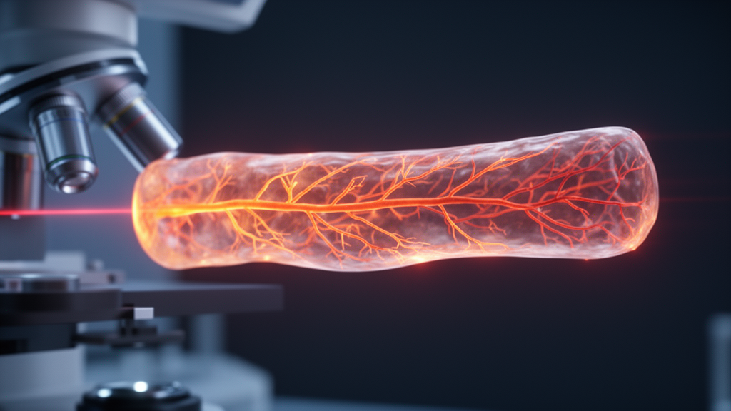
What the imaging showed was several signal-poor tubular structures within the adventitial layer in close association with graft side branches. These structures were visualized on serial cross-sectional and longitudinal images and extended longitudinally along the graft, and three-dimensional reconstruction demonstrated longitudinal continuity within the adventitial region. Their morphology and distribution were considered consistent with vasa vasorum. The authors note that the prominent tubular structure highlighted in their second figure was larger than the fine microvascular structures previously observed in no-touch grafts, and that its close association with a graft side branch and longitudinal course within the adventitial layer make it morphologically most compatible with a relatively proximal component of the vasa vasorum externa or a feeding vessel of that network. Vasa vasorum have been anatomically classified as vasa vasorum interna, vasa vasorum externa, and venous vasa vasorum, and additional serial FD-OCT images showed other adventitial tubuloluminal structures supporting the presence of vasa vasorum-related structures beyond the representative finding.
The limitations the authors acknowledge are substantial and worth stating plainly. FD-OCT provides morphological information only and cannot directly confirm blood flow, functional patency, or histological identity. Because pre-harvest or baseline imaging was not available, true preservation of pre-existing vasa vasorum could not be demonstrated. A residual side-branch remnant, another side-branch-related vascular structure, or a disconnected anatomical remnant could not be completely excluded. Although the persistence of these tubuloluminal structures two weeks after surgery may suggest that they were not immediately obliterated, the authors emphasize that this observation does not establish functional perfusion or biological activity, and it therefore remains uncertain whether these structures contributed to oxygen and nutrient supply of the graft wall.
Even with those caveats, the report carries a clear message for the field. In this case, only an approximately 1 to 2 millimeter rim of surrounding adipose tissue was retained, yet longitudinally continuous tubular structures were detected within the adventitial layer, suggesting that meticulous preservation of the adventitia may help retain adventitial structures consistent with vasa vasorum. The authors conclude that localized adventitial structures consistent with vasa vasorum were demonstrated in a conventional graft harvested with meticulous adventitial preservation, and that these findings are hypothesis-generating and require confirmation in comparative studies. A single case cannot establish whether such preservation translates into the superior patency associated with no-touch grafting, and the authors themselves point to the need for further comparative investigation of how preservation of adventitial structures affects graft patency and function. Until such studies are done, the report stands as a technical demonstration that modern intravascular optical imaging can resolve candidate vasa vasorum structures in living human vein grafts, and as an invitation to test whether refined conventional harvesting can capture some of the benefits long attributed to the no-touch technique.
Subject of Research: In vivo three-dimensional FD-OCT visualization of adventitial microvascular structures consistent with vasa vasorum in a conventionally harvested saphenous vein graft
Article Title: In vivo three-dimensional FD-OCT visualization of microvascular structures consistent with vasa vasorum in a conventionally harvested saphenous vein graft
Article References: Sugaya, A., Mizoguchi, M., Nakamura, M., & Kawahito, K. (2026). In vivo three-dimensional FD-OCT visualization of microvascular structures consistent with vasa vasorum in a conventionally harvested saphenous vein graft. General Thoracic and Cardiovascular Surgery Cases, 5(1), Article 39. https://doi.org/10.1186/s44215-026-00276-z
Image Credits: AI Generated
DOI: 10.1186/s44215-026-00276-z
Keywords: saphenous vein graft, vasa vasorum, coronary artery bypass grafting, FD-OCT, adventitia, no-touch harvesting, graft patency, three-dimensional reconstruction, case report, cardiac surgery, perivascular adipose tissue, optical coherence tomography
News Source: Ophelia Keating. (October 29, 2026). Optical imaging reveals vasa vasorum-like structures in conventionally harvested vein graft. Scienmag.