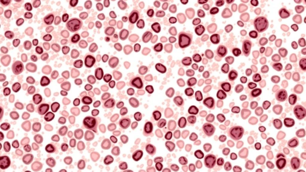
Among the many molecular and immunohistochemical markers that have swept through modern breast cancer diagnostics, two of the oldest and simplest measures of tumor cell proliferation remain at the center of an important debate: the manual mitotic count, in which a pathologist tallies dividing cells under the microscope, and Ki67, an immunohistochemical stain that labels cells at virtually every active phase of the cell cycle. A new population-based study from Norway, published in BMC Cancer, provides a rigorous head-to-head comparison of these markers and a third, antibody-based alternative, and arrives at a conclusion that could reshape how pathology laboratories classify and stratify the most common form of breast cancer. The research, led by Gøril Knutsvik and senior author Lars A. Akslen of the Centre for Cancer Biomarkers CCBIO at the University of Bergen, suggests that mitotic count may support or even replace Ki67 in separating aggressive from indolent luminal tumors.
The study addresses a genuinely practical clinical problem. Hormone receptor-positive, HER2-negative breast cancer accounts for the majority of breast cancer diagnoses worldwide, and within this broad category, tumors behave in strikingly different ways. Luminal A tumors tend to grow slowly and carry a favorable prognosis, while luminal B tumors proliferate faster and are more likely to recur. Distinguishing between the two is not an academic exercise: it directly influences whether patients receive chemotherapy in addition to endocrine therapy. Proliferation assessment is the linchpin of that distinction, and yet the field has never settled on a single, universally reliable way to measure it.
Mitotic count is baked into conventional histologic grading, where it contributes alongside tubule formation and nuclear atypia to the overall grade. However, the mitotic count component is not always reported as a separate value, which limits its visibility and utility in daily practice. Ki67, by contrast, is explicitly reported in many centers, but it suffers from a well-documented Achilles heel: a persistent lack of methodological standardization. Differences in antibody clones, staining protocols, scoring conventions, and the subjective choice of so-called hot spots have produced notorious interlaboratory variability, undermining confidence in any single cutoff value. The international working groups that have tried to harmonize Ki67 testing have made progress, but the reproducibility problem has never been fully solved.
The third marker examined in the study, phosphohistone H3 or PHH3, offers a technically elegant middle ground. PHH3 is an immunohistochemical antibody that specifically highlights cells in mitosis by staining a histone protein that becomes phosphorylated exclusively during mitotic chromosome condensation. In principle, PHH3 makes mitotic figures easier to spot against a background of crowded, atypical tumor cells, potentially reducing the subjectivity of manual counting while retaining the biological specificity of the mitotic count itself. The Norwegian team therefore compared all three proliferation measures—MC, Ki67, and PHH3—across different tissue preparation formats to see which combination delivered the most robust prognostic information.
That tissue-format question matters more than it might first appear. Whole sections (WS) represent the complete tumor as embedded in a surgical specimen, allowing pathologists to survey the entire lesion. Core needle biopsies (CNB), the tiny cylinders of tissue obtained during diagnostic puncture, offer only a limited sampling window. Tissue microarrays (TMA), in which minute cores from hundreds of tumors are arrayed on a single slide for high-throughput analysis, capture an even smaller snapshot of each tumor. Because proliferation can vary spatially within a tumor, the reliability of any proliferation marker could depend heavily on how much tissue is available for assessment. Whether markers validated on whole sections remain prognostic when measured on biopsies or microarray cores is therefore a question of direct relevance to modern, minimally invasive diagnostic pathways.
To answer it, the investigators turned to a prospective, population-based series of 534 women aged 50 to 69 years who were diagnosed with breast cancer between 1996 and 2003, with a median follow-up of 13 years. This long observation window is a major strength, because breast cancer recurrence and death can occur many years after diagnosis, and short follow-up periods can obscure prognostic signals. Mitotic count, Ki67, and PHH3-based mitotic counting were assessed in paired samples: whole sections from all 534 cases, core needle biopsies from 154 cases, and tissue microarray cores from 101 cases. In addition, the researchers compiled mitotic count data from four further independent cohorts comprising 1,598 cases to provide comparative context for their findings.
The results were consistent at the first level of analysis. In univariate survival analyses, all three proliferation markers showed a significant association with patient outcome, confirming that tumor cell proliferation, however it is measured, carries genuine prognostic weight in this population. The picture became more discriminating when the team applied multivariate statistical models, which adjust for other established prognostic factors and reveal whether a marker adds independent information. There, both mitotic count and Ki67 retained independent prognostic significance, demonstrating that each contributes something that the other clinical and pathological variables do not capture. PHH3, despite its theoretical appeal, did not sustain the same independent performance.
The most consequential finding emerged when the analysis was narrowed to the subgroup that matters most in current clinical decision-making: the hormone receptor-positive, HER2-negative, luminal tumors. This is precisely the patient group in which proliferation testing is used today to guide chemotherapy decisions. Within this subgroup, mitotic count retained its prognostic significance in multivariate analysis, whereas neither Ki67 nor PHH3 did. In other words, the humble count of mitotic figures, performed with a microscope and a calibrated field of view, outperformed the antibody-based proliferation indices in the very setting where such testing is most intensively applied. For a field that has invested heavily in standardizing Ki67, this is a provocative result.
The study also delivered a concrete translational number. In whole-section analysis of HER2-negative cases, a mitotic count of 2.5 mitoses per square millimeter corresponded to the widely used Ki67 cutoff of 20 percent, the conventional threshold for separating luminal B from luminal A tumors. This equivalence is technically meaningful because it anchors the newer, field-area-based method of reporting mitotic density to the established Ki67 framework that clinicians already understand. Reporting mitotic count per square millimeter, rather than per fixed number of high-power fields, also sidesteps a classic source of interlaboratory variation: microscope field sizes differ between instruments, and area-based counts remove that calibration dependence.
The authors conclude that manual proliferation markers are best assessed on whole sections for prognostication, a recommendation that carries implications for laboratories increasingly reliant on limited biopsy material and high-throughput microarray workflows. Their broader message is that mitotic count might support or even replace Ki67 as the stratification tool for luminal breast cancer, with a particular advantage in settings with limited resources. Unlike Ki67, mitotic counting requires no antibody, no staining platform, no external quality assurance scheme for immunohistochemistry, and no expensive reagents—only trained eyes and standardized methodology. In health systems where molecular assays and even reliable immunohistochemistry remain out of reach, a cheap, reproducible, and independently prognostic measure of proliferation could meaningfully improve risk stratification and treatment selection for the vast population of women with hormone receptor-positive breast cancer. The study, published open access with the DOI 10.1186/s12885-026-16925-z, invites pathology communities worldwide to reconsider whether the future of proliferation testing may lie, at least in part, in a technique as old as surgical pathology itself.
Subject of Research: Prognostic value of mitotic count, Ki67, and PHH3 proliferation markers in hormone receptor-positive breast cancer
Article Title: Tumor cell proliferation by mitotic count and Ki67 in prognostication and subclassification of hormone receptor-positive breast cancer
Article References: Knutsvik, G., Stefansson, I. M., Aziz, S., Klingen, T. A., Chen, Y., Arnes, J., Finne, K., Collett, K., Valla, M., & Akslen, L. A. (2026). Tumor cell proliferation by mitotic count and Ki67 in prognostication and subclassification of hormone receptor-positive breast cancer. BMC Cancer. https://doi.org/10.1186/s12885-026-16925-z
Image Credits: AI Generated
DOI: 10.1186/s12885-026-16925-z
Keywords: breast cancer, mitotic count, Ki67, PHH3, prognosis, luminal breast cancer, hormone receptor-positive, HER2-negative, histologic grade, tissue microarray, biomarkers, pathology
News Source: Nathaniel Bowman. (October 19, 2026). Old-Fashioned Mitotic Count May Outperform Ki67 in Hormone Receptor-Positive Breast Cancer. Scienmag.