Why Adult Plasticity Matters
Credit: Hedi Young
Why Adult Plasticity Matters
Much like young children who swiftly acquire languages in their early years, our visual system also has a “critical period” during the first few years of life where rapid development occurs. After this time, changes become more difficult, following the old adage, “You can’t teach an old dog new tricks”. Indeed, many treatments aimed at restoring vision, such as those addressing congenital cataracts or “lazy eye”, are only effective before the age of 7. With the advent of various established and emerging techniques to restore vision in adults, including gene therapy, bionic eyes, and surgeries, it’s vital to understand if the adult brain can even process new visual signals.
“If the adult brain lacks such plasticity or adaptability”, notes Noam Shemesh, the study’s senior author, “treatments targeting the eyes may prove futile if the brain is unable to interpret the incoming information. Interestingly, there are examples in nature, like birds rewiring their brains seasonally, or humans experiencing a brief window of plasticity after a stroke, which show that adaptation in adults is possible in certain circumstances”. The study’s central question, therefore, was to explore whether the adult mammalian brain still possesses the ability to reorganise its visual pathway and change even after the critical developmental period has passed.
Scientific and Technical Novelty
With the aid of a technical milestone, the researchers found that when rodents kept in the dark from birth were exposed to light for the first time in adulthood – long after the critical period has elapsed – their brains underwent significant reorganisation and adaptation, displaying a remarkable degree of plasticity. These findings not only provide evidence that the adult brain remains highly plastic, challenging previous beliefs about adult brain rigidity, but also open up new avenues for visual rehabilitation treatments.
As Noam Shemesh points out, the journey towards these revelations was fraught with technical hurdles. “Joana Carvalho, our lead researcher, faced numerous challenges and even doubts from some of the world’s leading labs who thought her endeavour was impossible. But Joana’s perseverance paid off. Without her resolve and creativity, we never would have reached this point. I really give Joana the credit for that”. Carvalho had to overcome the unprecedented difficulty of fitting a screen inside the constrained space of a rodent MRI scanner to project images onto it. “Due to space limitations and material constraints due to the ultrahigh magnetic field”, notes Carvalho, “previous studies in rodents only displayed flashes of light. Our method enables us to extract more detailed information compared to simple flashing visual stimuli”.
The Experiment
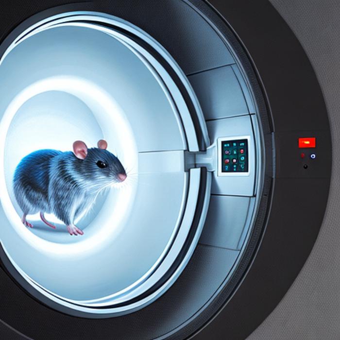
With their novel functional MRI (fMRI) setup, the team displayed intricate, patterned stimuli to the animals, and noninvasively mapped brain-wide properties previously accessible only through invasive techniques. “Initially”, Carvalho explains, “the challenge was to project images into a confined space riddled with obstructions, ensuring the mouse could view them without hindrance. The extremely high magnetic field of the MRI, capable of lifting a train, posed another substantial hurdle. We had to work around these constraints, using mirrors and specialised hardware, to get the images to where they needed to be. It helped that the rats were sedated, keeping their spontaneous eye movements and other motions to a minimum”.
Having surmounted these challenges, the researchers set out to explore the adult brain’s adaptability to visual signals. They used a model in which rodents are born and raised in the dark until adulthood, well past the critical period of plasticity. Consequently, the brains of these animals had not yet undergone the key processes required for visual specialisation. The animals were then exposed to light for the first time inside the MRI scanner. This allowed the researchers to not only observe the brain’s response to its first encounter with visual stimuli, but also to study how it might adapt to this delayed exposure, yielding two pivotal insights.
First, when the animals were exposed to light for the first time during the initial MRI scan, their brains displayed no organised response to visual information. Instead, their nerve cells across different areas reacted to a broad range of visual details, from fine to coarse. Moreover, the receptive field sizes of neurons – the specific area of the visual field that they respond to – was also larger in visually deprived rats compared to the control group. Together, these findings suggested that the visual pathway in the light-deprived rats lacked specialisation.
Second, after exposure to light, the animals’ brains began to change. Even within a week, visual responses became more organised, such that neighbouring neurons began to respond to nearby positions in the visual field, and the cells started to react more to specific visual characteristics. The receptive fields of the neurons also became smaller and more spatially selective. After a month, the animals’ brains looked much like those of healthy controls.
“Surprisingly”, says Shemesh, “in less than a month, the structure and function of the visual system in the visually deprived animals became similar to the controls. While plasticity has been observed in humans, interpreting it remains very difficult. What we are seeing here in rodents, which offer insights into brain mechanisms unattainable in human studies, is a phenomenon that has not been observed before: large-scale plasticity in the adult brain across the entire visual pathway, not just localised to a specific brain area as shown in previous papers”.
Prior studies had used electrophysiology and calcium imaging, which focus on isolated brain regions, and lack a comprehensive view of the entire pathway. These methods – while providing direct readouts of neural activity – are invasive, potentially introducing confounds, and the difficulty of monitoring the same cells at different times with these techniques may lead to detecting changes unrelated to actual plasticity.
While lacking single cell specificity and indirectly reflecting neuronal activity, fMRI facilitates the longitudinal and non-invasive measurement of entire visual areas simultaneously with very high resolution. “As a result, one of the intriguing things we were able to notice”, reveals Carvalho, “was that a part of the visual pathway called the superior colliculus seemed to take longer to adapt in visually deprived animals compared to other areas, like the cortex. It’s something we’d love to explore further. This also highlights the importance of an integrative view of the entire system in the same animal over multiple time points”.
Potential clinical implications and looking ahead
“We’re now in a position to start exploring whether we can predict which animals may have improved or deteriorated vision based on the MRI responses of their visual system”, remarks Shemesh. “In animals with impaired vision, we’d like to determine which ones will benefit most from certain therapeutic interventions. Currently, it’s challenging for medical doctors to determine from an MRI scan whether a patient’s brain will respond to a particular treatment, leading to unnecessary suffering and lost time. Through preclinical imaging, we can begin to chart treatment responses in rats, which could not only deepen our understanding of the treatment’s effects but also accelerate the pace of treatment development in humans, as well as guide clinicians on the necessary scans for their patients”.
Furthermore, the techniques from this study are extendable to other animal disease models, including, for example, Parkinson’s Disease, which is also being studied in the Shemesh Lab. As there are known early, subtle visual problems in Parkinson’s, the method could be applied to track differences in visual system responses over time, possibly revealing new insights into disease progression and treatment options in animal models. Adds Shemesh, “within the preclinical setting, this technique could assist in pinpointing the optimal timing for visual restoration and rehabilitation procedures, enhancing the effectiveness of treatments like retinal stem cell transplantation”.
Meanwhile, the team continues to forge ahead. Carvalho is keen to explore the neural mechanisms that drive the adaptation of the visual system in light-deprived rats, in particular focusing on excitatory–inhibitory balances and the role of long-range connections. Shemesh intends to build on Carvalho’s innovations to conduct experiments in awake, non-sedated rats, which will require overcoming further challenges, such as prolonged training to acclimate the animals to scanner noises and to maintain a fixed gaze to avoid eye movement-induced distortions. The Champalimaud Foundation’s acquisition of an 18 Tesla MRI scanner, the most powerful horizontal scanner in the world, will no doubt facilitate their efforts to understand and enhance plasticity in adult humans, and perhaps one day, even in old dogs too.
Journal
PLoS Biology
DOI
10.1371/journal.pbio.3002229
Method of Research
Experimental study
Subject of Research
Animals
Article Title
Extensive topographic remapping and functional sharpening in the adult rat visual pathway upon first visual experience
Article Publication Date
17-Aug-2023

