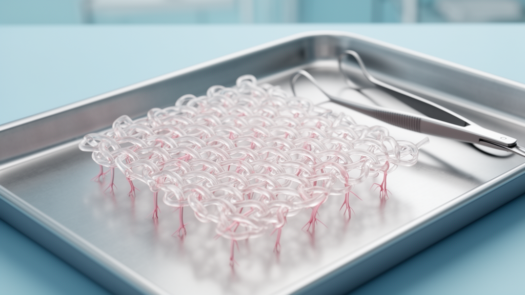
A novel hybrid knitted patch designed to invite a patient’s own tissue to grow into it has been examined for the first time in a human infant, and the histological findings are drawing attention for what they reveal about early healing around cardiovascular implants. In a case report published in General Thoracic and Cardiovascular Surgery Cases, surgeons and researchers in Japan describe removing the patch ten months after it was used to enlarge the right ventricular outflow tract of a one-month-old baby with pulmonary atresia and an intact ventricular septum. Under the microscope, both surfaces of the explanted fabric were covered with regenerated tissue containing smooth muscle cells, blood vessels, and collagen fibers, with only a modest inflammatory response.
The material at the center of the report, known commercially as SYNFOLIUM, was developed as an alternative to the patch materials surgeons currently reach for when reconstructing the right ventricular outflow tract, the muscular corridor that carries blood from the right ventricle toward the lungs. Existing options, including autologous or xenograft pericardium, homografts, bovine jugular vein, expanded polytetrafluoroethylene, and various extracellular matrix products, share two persistent shortcomings: they lack growth potential, and they are prone to calcification over the long term. In pediatric cardiac surgery, where the patch must ideally grow along with the child, those limitations complicate the postoperative course.
The hybrid fabric is knitted from a combination of bioabsorbable poly L-lactic acid yarns and nonabsorbable polyethylene terephthalate yarns, and it is coated with cross-linked gelatin. Once implanted, the bioabsorbable component is designed to degrade gradually, opening space for autologous tissue to grow into the knit. The remaining nonabsorbable knit retains a stretchable structure intended to accommodate the patient’s physical growth. According to the authors, this process of in situ tissue regeneration has the potential to mitigate the inflammatory and foreign body reactions, and the cell death, that lead to implant deterioration. The material received regulatory approval in Japan on July 12, 2024, and has since been used in both pediatric and adult patients with congenital heart disease.
The clinical story behind the explant began before birth. The infant was born at 38 weeks with a birth weight of 3.1 kilograms and was placed on a prostaglandin E1 infusion to keep the fetal ductus arteriosus open. Balloon atrial septostomy was performed on the tenth day after birth. Right ventricular angiography revealed a tripartite right ventricle with an end-diastolic volume of 4 milliliters, 44 percent of normal, and echocardiography showed severe tricuspid regurgitation despite a nearly normal-sized tricuspid valve annulus.
On the 38th postnatal day, the surgical team performed an open pulmonary valvotomy, enlargement of the right ventricular outflow tract, partial closure of the atrial septal defect, and ligation of the ductus arteriosus, working through a median sternotomy on cardiopulmonary bypass. The atretic pulmonary valve proved to be thin, with three fused commissures and well-balanced leaflets, leading the surgeons to predict that the native valve could function adequately if the annulus developed properly after the valvotomy. After sharp incisions along the fused commissures, a 5 millimeter Hegar dilator passed smoothly through the valve orifice. The infundibulum of the right ventricle was then incised just beneath the pulmonary valve, the parietal band was partially resected, and a trimmed hybrid wrap-knitted fabric patch was sutured to the incision with continuous monofilament suture to enlarge the outflow tract. The patient was discharged 41 days after surgery in stable condition.
The patch itself performed without structural incident, but the native valve did not. Eight months after the operation, recurrent severe pulmonary valve stenosis developed, with an echocardiographic peak flow velocity of 5.5 meters per second and a catheterization-derived pressure gradient of 135 millimeters of mercury. Right ventricular angiography showed the patch-covered segment of the outflow tract distending immediately proximal to the stenotic pulmonary valve annulus and the narrowed infundibulum. At ten months of age, the infant underwent repeat reconstruction of the outflow tract, during which the hybrid patch was replaced with a handmade tri-leaflet expanded polytetrafluoroethylene conduit. The second operation also provided the opportunity the researchers had been waiting for: a close look at how the novel fabric had behaved inside a living, growing heart.
Grossly, the explanted patch showed no thinning, aneurysmal dilation, or structural failure, even though it had functioned under right ventricular pressures exceeding systemic levels. Both surfaces were covered with autologous tissue, although that tissue could be easily detached from the fabric. Histological examination added detail to this picture. Collagen fiber-rich tissue had developed around the patch on its proximal right ventricular side, and despite a modest infiltration of neutrophils and lymphocytes, there was no significant chronic inflammation or foreign body reaction to the patch material. Throughout the gelatin absorption process, the structural arrangement of all the yarns was preserved. Immunohistochemistry revealed smooth muscle cells and small-caliber vasculature within the regenerated tissue.
Transmission electron microscopy pushed the analysis to the cellular scale. Cardiomyocytes were absent from the tissues surrounding the patch, but histiocytic granulomas containing macrophages were observed adjacent to the patch material. The tissue also exhibited thick-walled capillaries lined with endothelial cells, along with neutrophil infiltration. Most strikingly, the sample contained mature fibroblasts with dilated rough endoplasmic reticulum and an increased number of collagen fibers, a profile consistent with active, collagen-producing cells. The authors interpret this cellular constellation as evidence of potential macrophage-fibroblast interaction promoting tissue remodeling within the new fabric, resembling a local migratory response to myocardial injury that drives remodeling.
The authors frame two key findings from this first human look at the material’s early biology. First, the patch functioned without complication under pressures exceeding systemic levels, and histology confirmed no aneurysmal changes, which they suggest may have been prevented by the maintained arrangement of all fibers and their attachment to the gelatin coating. They note this demonstration of safety under systemic pressure had not been reported in the previous clinical trial. Second, the autologous tissue formed on the patch surface contained smooth muscle cells, microvessels, and collagen fibers with only modest inflammation, suggesting tissue restoration or regeneration. That contrasts with previous reports on decellularized xenogeneic patches, whose surface lining is frequently a relatively avascular, densely fibrotic pannus with sustained chronic inflammatory infiltration and calcific degeneration, and with expanded polytetrafluoroethylene, which typically elicits a thin, largely acellular fibrous capsule with limited cellular or vascular ingrowth.
Important caveats temper the enthusiasm. Gelatin absorption in this patient was slower than observed in preclinical studies, and the authors suggest this delayed absorption may have impeded formation of bridging tissue, explaining why the autologous tissue detached so easily. They also conclude that the recurrent stenosis in this case appeared to result predominantly from the stenotic pulmonary valve annulus rather than from degeneration of the patch itself. Crucially, the findings support the material’s safety and biological performance but do not establish its efficacy in preventing reintervention; confirming long-term efficacy and safety for tissue restoration or regeneration will require histological analysis of clinical samples obtained after complete degradation of the bioabsorbable components, in larger and longer-term clinical series. For now, the report stands as an early, detailed portrait of how a regenerative knit behaves in the smallest and most demanding of patients.
Subject of Research: Early histological reactions to a novel hybrid knit cardiovascular patch implanted in an infant
Article Title: Early biological reactions to a novel hybrid knit fabric inducing autologous tissue ingrowth implanted in a small infant
Article References: Kido, T., Nemoto, S., Narita, J., Ishida, H., Ishii, R., Ueno, T., & Miyagawa, S. (2026). Early biological reactions to a novel hybrid knit fabric inducing autologous tissue ingrowth implanted in a small infant. General Thoracic and Cardiovascular Surgery Cases, 5(1), Article 38. https://doi.org/10.1186/s44215-026-00275-0
Image Credits: AI Generated
DOI: 10.1186/s44215-026-00275-0
Keywords: pulmonary atresia, right ventricular outflow tract, hybrid knit fabric, tissue ingrowth, pediatric cardiac surgery, SYNFOLIUM, patch explantation, histology, macrophage-fibroblast interaction, gelatin coating, PLLA, case report
News Source: Ophelia Keating. (October 29, 2026). Novel Knitted Heart Patch Shows Early Tissue Ingrowth in Infant Case Report. Scienmag.