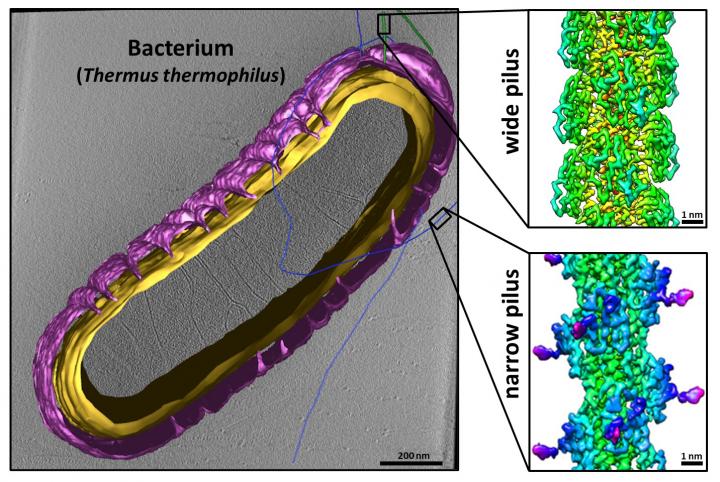
Credit: University of Exeter
Scientists have made a pivotal breakthrough in advancing our understanding of how bacteria move and perform genetic exchange – that could potentially lead to the development of new antimicrobial drugs.
A team of researchers from the University of Exeter’s Living Systems Institute and the University of Frankfurt has made a crucial discovery around the structures of long filaments – hair like appendages – called type IV pili found on the surface of bacteria.
Type IV pili are known to play an important role in how bacteria proliferate and form biofilms, through movement, genetic exchange, adhesion and communicating with other cells.
Genetic exchange occurs when cells take up DNA – the molecule that encodes an organisms’ genetic code – from their environment.
DNA transfer plays a vital role in the bacteria’s ability to become resistant to treatments. There are antibiotic resistance genes, for example, that get shared and thus render the treatment useless.
In this new research, scientists have discovered that the bacterium Thermus thermophilus can produce two types of type IV pili – one specialised for movement and one for genetic exchange.
The pioneering research could allow scientists to target the two functions independently, for example by developing new drugs that stop bacteria from moving or becoming resistant to antibiotics.
The study is published in leading journal Nature Communications on Wednesday, May 6th 2020.
Dr Vicki Gold, a Senior Lecturer at the University of Exeter and lead author of the paper said: “It will now be important to investigate if this phenomenon is a universal principle occurring in other bacteria expressing type IV pili. This would pave the way for the development of antimicrobials aimed to target a particular mechanism.”
Type IV pili are protrusions that are found across the surface of a bacterial cell, and are made up of thousands of copies of one protein.
In the new study, the researchers used cryo-electron microscopy to determine structures of both type IV pili in unprecedented detail. The technique allowed the researchers to gather a vast array of detailed images of the structures in different orientations to create a detailed, 3D picture.
The discovery that one of the pili is composed of a previously uncharacterised protein means that scientists are able to target the different functions to determine what is important for microbial proliferation and genetic exchange.
As a result, they are able to conduct experiments to see how well bacteria proliferate, or take up DNA, when certain type of pilus formation are artificially impeded.
Alexander Neuhaus, first author of the research and also from the University of Exeter said: “It is going to be interesting to discover how exactly the two pili fulfil their different functions and how bacteria control their production. Knowledge of these mechanisms could lead to new strategies for combatting bacterial infection.”
###
Cryo-electron microscopy reveals two distinct type IV pili assembled by the same bacterium is published in Nature Communications on Wednesday, May 6th 2020.
The research was carried out in collaboration with the laboratory of Prof. Beate Averhoff (University of Frankfurt) and is funded by the BBSRC.
Media Contact
Duncan Sandes
[email protected]
Related Journal Article
http://dx.


