Bone is the most common site of distant metastasis in breast cancer, with up to 75 percent of patients with advanced disease developing skeletal involvement, and it is the third most common metastatic site across all cancer types. Yet studying how cancer cells actually behave once they reach bone has remained one of the most stubborn obstacles in late-stage cancer research, because the standard laboratory tools each fall short in different ways. A team at the University of Notre Dame has now published a detailed protocol for a new ex vivo co-culture system that keeps intact mouse bone and its marrow alive in a dish for weeks while breast cancer cells colonize it, offering researchers a middle ground between oversimplified cell culture and hard-to-control animal experiments.
The work, published in BMC Methods by Ricardo J. Romero-Moreno, Courtney L. Flatt, Madeline P. Sheeley, Xiaotong Alex Lin, Glen L. Niebur and Laurie E. Littlepage, describes a step-by-step procedure for extracting femurs and tibiae from mice, injecting cancer cells directly into the bone marrow cavity, and maintaining the resulting co-culture for extended periods. The system preserves viable and metabolically active bone, marrow and cancer cells for up to four weeks and retains the heterogeneity of the native bone microenvironment, something that engineered alternatives such as hydroxyapatite nanoparticles, biomimetic collagen hydrogels, bone-on-a-chip devices and microfluidic platforms have struggled to fully reproduce.
The motivation behind the method lies in a clinical problem that has long frustrated oncologists. Metastasis is the cause of most breast cancer-related deaths, yet many therapies are designed to treat the primary tumor even though metastases often do not respond to the same treatments. As cancer cells adapt to new environments during metastatic spread, they undergo significant morphological and physiological changes. By the time cells have colonized distant tissue, they frequently lack the biomarkers that guided therapy in the primary tumor: expression of key targets including estrogen receptor, progesterone receptor and HER2 often differs between primary breast tumors and bone metastases. That leaves many patients with aggressive chemotherapy as their remaining option, which is why the authors argue it is imperative to study the direct interactions between cancer cells and their secondary tissue host.
In vitro models, typically flat monolayers of cells, do not mimic the three-dimensional structure or cellular diversity of intact bone, making results difficult to translate to more complex settings. In vivo models, meanwhile, are difficult to control for specific variables given the complexity of an entire organism, and metastasis experiments in animals can be confounded by the primary tumor burden, sometimes forcing premature sacrifice of the animals and compromising the interpretation of the data. The new protocol was adapted from earlier work on culturing trabecular bone cores from large mammals, modified to keep whole mouse bones intact and then seeded with mouse breast epithelial cancer cells before the culture period begins.
The procedure itself is deliberately practical. Bone extraction takes roughly one and a half to two hours, and the researchers advise working quickly, since success depends heavily on prompt removal of bones from the mice and immediate transfer into pre-warmed culture media to minimize cell death. Each femur and tibia is cleaned of muscle and connective tissue, its cartilage caps removed, and holes drilled or cut at both ends to expose the marrow cavity so nutrients can diffuse in. Each bone is then bisected at the midshaft, and half a femur and half a tibia are placed together in a single well of a low-binding 12-well plate containing 2 milliliters of high-glucose DMEM supplemented with 10 percent fetal bovine serum and 1 percent penicillin-streptomycin. The full setup can be completed in a single day, and daily maintenance thereafter takes only about 15 minutes.
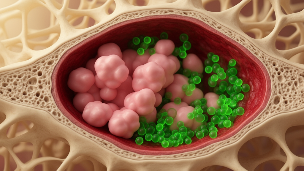
Cancer cells, kept at around 80 percent confluence, are trypsinized, counted and resuspended at 2.5 times ten to the fifth cells per 10 microliters of media, then injected into the marrow cavity at the cut midshaft end of each explant using a media-coated 27-gauge needle. A larger injection volume risks flushing the marrow out, the authors warn. The use of anchorage-dependent cancer cells grown in low-attachment plates is a deliberate design feature: the cells must be in contact with the bone to survive, ensuring that any growth observed reflects genuine colonization of the bone environment rather than independent proliferation in suspension. Control wells include plain media without bone, bones receiving mock injections of phosphate-buffered saline, and devitalized bone that has been through at least five freeze-thaw cycles.
Once established, the co-culture supports several functional readouts. If the cancer cells carry a luciferase reporter gene, as the PyMT mouse mammary tumor line used in the protocol does, bioluminescence imaging can track cancer cell proliferation and metabolic viability throughout the experiment: D-luciferin is spiked into each well to a final concentration of 150 micrograms per milliliter, cultures incubate for 10 minutes, and luminescence is measured on a plate reader or IVIS imaging system. Daily partial media changes also allow conditioned media to be collected, flash-frozen and stored for analysis of secreted factors. In the published validation experiments, the bone remodeling factors RANKL and OPG were both detectable by ELISA in conditioned media collected on day 5 of culture, demonstrating that bone cell signaling remains active in the explants.
Viability and structural integrity hold up remarkably well over the culture period. Using the CellTiter-Blue reagent, which converts resazurin to fluorescent resorufin, the team showed that cultured bones maintained significantly higher metabolic activity than dead bone controls for up to four weeks. Immunohistochemistry of co-cultured bones revealed that a broad range of immune and bone cells, including CD4-positive helper T cells, CD8-positive cytotoxic T cells, CD20-positive B cells, CD68-positive macrophages, Ly6G/6C-positive neutrophils, endomucin-positive endothelial cells, CD61-positive megakaryocytes and CD71-positive erythroid precursors, remained alive and metabolically active after two weeks. Micro-computed tomography confirmed that bone volume fraction and trabecular architecture in the proximal tibia of cultured bones after two weeks were preserved compared with fresh, uncultured bones, and µCT scanning must be completed before decalcification while bones sit in 70 percent ethanol.
After the culture period, bones are fixed overnight in 4 percent paraformaldehyde, decalcified in 10 percent EDTA at 4 degrees Celsius for one to three weeks with weekly solution changes, then paraffin-embedded and sectioned at 4 to 6 micrometers for immunostaining. The authors note trade-offs in decalcification: higher-temperature decalcification at 37 degrees Celsius shortens the process considerably but increases background staining for immunohistochemistry and immunofluorescence, though chemical stains such as hematoxylin and eosin are largely unaffected. A detailed troubleshooting section addresses common failure modes, from bone and marrow cell death caused by insufficient nutrient perfusion, to failed cancer cell injections, to loss of secreted factors through improper handling and freeze-thaw cycles, to sectioning problems caused by under-decalcification.
The platform does have defined limits. It was developed and optimized with PyMT mouse mammary epithelial cancer cells in bones from FVB/N mice, and other cell lines have not yet been validated. In early experiments, human MDA-MB-231 breast cancer cells co-cultured with the same FVB/N mouse bones did not achieve the same level of survival as the mouse line, which the authors suggest may reflect an immune response from the mouse marrow against human cells; further testing with syngeneic or immunocompromised mice will be needed. Cell lines not known to metastasize to bone have not been tested, and researchers using different media requirements must separately verify that both cancer cells and bone cells tolerate the conditions. The protocol requires basic expertise in mammalian cell culture and mouse dissection, and the authors recommend additional personnel if more than six mice are processed at once, since extended processing times compromise bone and marrow viability. Within those boundaries, the system offers an adaptable platform for studying tumor-stroma interactions, testing treatments against cancer cells inside a living bone environment, and potentially supporting preclinical therapeutic screening in the bone metastatic niche.
Subject of Research: An ex vivo co-culture protocol for modeling breast cancer metastasis to bone using mouse long bone explants
Article Title: A novel bone and cancer cells co-culture methodology to model breast cancer metastasis to bone ex vivo
Article References: Romero-Moreno, R. J., Flatt, C. L., Sheeley, M. P., Lin, X. A., Niebur, G. L., & Littlepage, L. E. (2026). A novel bone and cancer cells co-culture methodology to model breast cancer metastasis to bone ex vivo. BMC Methods, 3(1), Article 36. https://doi.org/10.1186/s44330-026-00085-4
Image Credits: AI Generated
DOI: 10.1186/s44330-026-00085-4
Keywords: bone metastasis, breast cancer, ex vivo culture, co-culture, bioluminescence imaging, micro-computed tomography, tumor microenvironment, mouse model, cancer research, protocol, bone marrow, preclinical screening
News Source: Nathaniel Bowman. (October 28, 2026). New Ex Vivo Co-Culture Method Grows Breast Cancer Cells Inside Living Mouse Bone. Scienmag.