• HOME
  • NEWS
  • EXPLORE
    • CAREER
      • Companies
      • Jobs
    • EVENTS
    • iGEM
      • News
      • Team
    • PHOTOS
    • VIDEO
    • WIKI
  • BLOG
  • COMMUNITY
    • FACEBOOK
    • INSTAGRAM
    • TWITTER
Sunday, October 4, 2026
BIOENGINEER.ORG
No Result
View All Result
  • Login
  • HOME
  • NEWS
  • EXPLORE
    • CAREER
      • Companies
      • Jobs
        • Lecturer
        • PhD Studentship
        • Postdoc
        • Research Assistant
    • EVENTS
    • iGEM
      • News
      • Team
    • PHOTOS
    • VIDEO
    • WIKI
  • BLOG
  • COMMUNITY
    • FACEBOOK
    • INSTAGRAM
    • TWITTER
  • HOME
  • NEWS
  • EXPLORE
    • CAREER
      • Companies
      • Jobs
        • Lecturer
        • PhD Studentship
        • Postdoc
        • Research Assistant
    • EVENTS
    • iGEM
      • News
      • Team
    • PHOTOS
    • VIDEO
    • WIKI
  • BLOG
  • COMMUNITY
    • FACEBOOK
    • INSTAGRAM
    • TWITTER
No Result
View All Result
Bioengineer.org
No Result
View All Result
Home NEWS Science News Biology

New cable-free brain imaging method may take social neuroscience to the next level

Bioengineer by Bioengineer
May 28, 2019
in Biology
Reading Time: 3 mins read
0
IMAGE
Share on FacebookShare on TwitterShare on LinkedinShare on RedditShare on Telegram

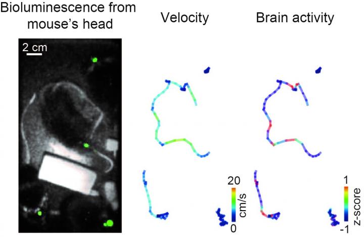
Researchers at Osaka University develop a cable-free recording method that can be used to measure brain activity associated with social behavior in mice
IMAGE

Credit: Professor Takeharu Nagai

Osaka, Japan – Existing electrophysiological and fluorescence-based brain imaging techniques in mice are generally invasive, require head fixes or cables, and are not suitable for long-term recordings. While there have been recent advances in imaging methods in freely moving animals, these are major limitations for researchers that are interested in the brain correlates of social behaviors.

Researchers at Osaka University have developed a new method to record brain activity simultaneously in multiple, freely moving mice. The method is based on a recent bioluminescence-based indicator of membrane voltage called “LOTUS-V”. The LOTUS-V bioluminescent probe is genetically encoded, which means that it is delivered to target cells non-invasively via a common gene expression system (the adeno-associated virus). Its signal is derived from cell membrane voltage changes, which reflect brain activity.

“The LOTUS-V method reported brain activity in freely moving mice with a good sensitivity and without motion artifacts,” says corresponding author Takeharu Nagai. “More importantly, it could measure dynamically changing brain activity in the primary visual cortex during social interactions.”

LOTUS-V was applied to cells in the primary visual cortex; this area was targeted because it is known to respond to locomotor activity and visual stimulation. LOTUS-V signal changes reflected neural activity in response to visual stimuli and locomotor activity, as well as during interactions with other mice; neural activity was significantly higher when a mouse approached others. Furthermore, the LOTUS-V signal was not affected by leaky signals emitted from other, nearby mice, which means that it faithfully reflected in vivo brain activity.

“Our method successfully detected activity of the superficial layer of the primary visual cortex–this is about 300 μm deep,” says Shigenori Inagaki, first author of the study. “It will be important to test its applicability to recording in deeper brain regions.”

While the temporal resolution of the LOTUS-V method was sufficient to investigate the dynamics of brain activity triggered by specific events, it is not yet superior to that of the fiber-based method.

“These results could be really exciting for social neurobiologists,” Nagai says. “It is minimally invasive, doesn’t require cables or head fixes, and is suitable for long-term recordings in freely moving animals, meaning it could be useful in a broad range of other research fields, too.”

###

The article, “Imaging local brain activity of multiple freely moving mice sharing the same environment”, was published in Scientific Reports at DOI: https://doi.org/10.1038/s41598-019-43897-x

About Osaka University

Osaka University was founded in 1931 as one of the seven imperial universities of Japan and now has expanded to one of Japan’s leading comprehensive universities. The University has now embarked on open research revolution from a position as Japan’s most innovative university and among the most innovative institutions in the world according to Reuters 2015 Top 100 Innovative Universities and the Nature Index Innovation 2017. The university’s ability to innovate from the stage of fundamental research through the creation of useful technology with economic impact stems from its broad disciplinary spectrum.

Website: https://resou.osaka-u.ac.jp/en/top

Media Contact
Saori Obayashi
[email protected]

Related Journal Article

http://dx.doi.org/10.1038/s41598-019-43897-x

Tags: BiologySocial/Behavioral ScienceTechnology/Engineering/Computer Science
Share12Tweet8Share2ShareShareShare2

Related Posts

Hidden Genome: Tiny Proteins Mapped Across the Parasite Behind Trichomoniasis

Hidden Genome: Tiny Proteins Mapped Across the Parasite Behind Trichomoniasis

October 4, 2026
Mint Compound L-Carvone Shows Promise Against Uremia-Driven Blood Vessel Damage in Kidney Disease

Mint Compound L-Carvone Shows Promise Against Uremia-Driven Blood Vessel Damage in Kidney Disease

October 4, 2026

Why Starch Never Predicts Texture Alone: The Matrix Rules That Decide How Food Feels

October 4, 2026

Gene Activity Comes in Bursts: Epigenetics Reshapes the Picture of Transcriptional Noise

October 4, 2026
Please login to join discussion

POPULAR NEWS

  • Alloys That Shrink Their Own Grains: New PIX Mechanism Refines Metals With Heat Alone

    Alloys That Shrink Their Own Grains: New PIX Mechanism Refines Metals With Heat Alone

    29 shares
    Share 12 Tweet 7
  • Endurance Exercise Reshapes the Liver in Males and Females Through Distinct Molecular Routes

    29 shares
    Share 12 Tweet 7
  • Single Transcription Factor PU.1 Rapidly Converts Fibroblasts into Macrophage-Lineage Cells

    29 shares
    Share 12 Tweet 7
  • New Scale Measures How Ready Nurse Educators Really Are for the AI Era

    29 shares
    Share 12 Tweet 7

About

We bring you the latest biotechnology news from best research centers and universities around the world. Check our website.

Follow us

Recent News

Alloys That Shrink Their Own Grains: New PIX Mechanism Refines Metals With Heat Alone

Endurance Exercise Reshapes the Liver in Males and Females Through Distinct Molecular Routes

Single Transcription Factor PU.1 Rapidly Converts Fibroblasts into Macrophage-Lineage Cells

Subscribe to Blog via Email

Enter your email address to subscribe to this blog and receive notifications of new posts by email.

Join 85 other subscribers
  • Contact Us

Bioengineer.org © Copyright 2023 All Rights Reserved.

Welcome Back!

Login to your account below

Forgotten Password?

Retrieve your password

Please enter your username or email address to reset your password.

Log In
No Result
View All Result
  • Homepages
    • Home Page 1
    • Home Page 2
  • News
  • National
  • Business
  • Health
  • Lifestyle
  • Science

Bioengineer.org © Copyright 2023 All Rights Reserved.