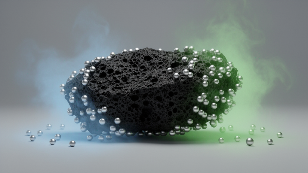
Silver ions are among the more troublesome contaminants in water: useful in industry for their antimicrobial and conductive properties, yet toxic and highly mobile once released into ecosystems. A new open-access study published in Biochar on 26 August 2026 reports that a carefully engineered form of nano-biochar can convert dissolved silver ions into silver nanoparticles, but only within a narrow alkaline window, and that superoxide radicals are the decisive chemical intermediates making the transformation happen.
The research, led by Shiguo Gu, Dandan Wang, Tongtong Wang and Wei Zhu, centered on nano-biochar particles smaller than 100 nanometers, produced by ball milling bulk biochar derived from wood chips. The team prepared two versions: one from biochar pyrolyzed at 400 degrees Celsius, labeled nano400, and another from biochar pyrolyzed at 700 degrees Celsius, labeled nano700. This temperature contrast was deliberate, because heat treatment strongly shapes the surface functional groups, persistent free radicals and electron-donating capacity that the particles carry into a reaction.
When the researchers suspended the nano-biochar in a 0.5 millimolar silver nitrate solution and adjusted the pH across a range from 6.5 to 10, a striking pattern emerged. At acidic to neutral pH values between 6.5 and 7.5, no silver nanoparticles formed at all. Once the solution was shifted to weakly alkaline conditions between pH 8.5 and 9.5, reduction efficiency improved markedly, and the highest yield of silver nanoparticles appeared at pH 9.5. The team attributes the acidic failure to protonation of the biochar’s surface functional groups, which leaves too few reactive sites and too little electron-donating capacity to drive the reaction forward.
Characterization work helped explain why the two biochar types behaved so differently. X-ray photoelectron spectroscopy showed that nano700 carried a higher share of carbon-carbon bonds, consistent with the graphene-like structures that form at high pyrolysis temperatures, while nano400 was richer in oxygen-containing functionalities, including hydroxyl and carbonyl groups. Boehm titration put the phenolic hydroxyl content of nano400 at 3.01 millimoles per gram, roughly 2.66 times the 1.13 millimoles per gram measured for nano700. Nano400 also showed a higher absolute zeta potential across the tested pH range, which favors both colloidal stability and electrostatic adsorption of positively charged silver ions onto the particle surface.
Kinetic analysis reinforced the advantage of the lower-temperature material. Formation of silver nanoparticles followed pseudo-first-order kinetics, and nano400 posted a reaction rate constant approximately 2.5 times higher than that of nano700 at pH 9.5. The authors connect this directly to structure: nano400’s 2.66-fold higher phenolic hydroxyl content matched its 2.50-fold higher formation rate constant, which they present as quantitative evidence that phenolic hydroxyl groups are the dominant electron-donating structural determinants of the reduction kinetics. Electrochemical measurements showed nano400’s intrinsic electron-donating capacity was about 1.56 times that of nano700.
The mechanistic core of the study lies in the radicals. Using probe molecules, superoxide dismutase quenching assays and electron paramagnetic resonance spectroscopy with DMPO spin trapping, the team identified superoxide radicals as the key intermediates. At pH 9.5, nano400 produced 1.56-fold higher XTT formazan levels than nano700, and DMPO-superoxide signals appeared across the pH 8.5 to 9.5 range, growing with pH. When superoxide dismutase was added to quench superoxide, nanoparticle formation was inhibited, and anoxic, nitrogen-saturated control experiments produced no silver nanoparticles at all, indicating that direct electron transfer from deprotonated phenolic groups to silver ions was negligible.
According to the proposed pathway, persistent free radicals on the nano-biochar surface, formed by homolytic cleavage of phenolic hydroxyl groups during pyrolysis and stabilized by sp2 carbon conjugation, donate electrons to dissolved oxygen to generate superoxide. The biochar’s reduction potential, reported between minus 0.36 and minus 0.49 volts versus Ag/AgCl, is low enough to mediate the reduction of oxygen to superoxide, which sits at minus 0.28 volts. Adsorption of silver ions onto the deprotonated, negatively charged biochar surface, maximized at pH 9.5, then positions them for reduction by the interfacial superoxide. The authors describe this as a catalytic cycle in which phenolic groups donate initial electrons while dissolved oxygen continuously regenerates superoxide, sustaining the reaction without net consumption of the biochar’s active sites.
Dosage proved to be a double-edged variable. Moderate loadings of nano-biochar accelerated the reaction, but excessive amounts inhibited silver nanoparticle formation, with the threshold sitting above 10 milligrams per liter for nano400 and above 20 milligrams per liter for nano700. The authors attribute this inhibition to reduced interfacial reactive sites and self-consumption of reactive oxygen species, noting that disproportionation of superoxide into singlet oxygen and hydrogen peroxide, and hydroxyl radical production, increase as biochar content rises.
The nanoparticles produced under optimal conditions were characterized in detail. Transmission electron microscopy revealed quasi-spherical particles with a narrow size distribution between 5.88 and 23.53 nanometers, well dispersed during the 10-minute reaction. High-resolution TEM identified lattice fringes corresponding to the {111}, {200} and {220} planes of face-centered cubic silver, and selected area electron diffraction confirmed a polycrystalline structure. Energy dispersive spectroscopy detected a prominent silver emission peak, and a color transition of the suspensions from pale yellow to deep yellow with rising pH provided a visible confirmation of nanoparticle formation. A control group without biochar showed no measurable nanoparticle generation throughout the reaction period.
The authors frame the work as theoretical support for nano-biochar-based remediation of silver-contaminated water, noting that converting the more toxic ionic form into nanoparticles could mitigate aquatic ecological risks, since prior studies cited in the paper report that silver ions are taken up by plants 67 to 87 times faster than silver nanoparticles and show greater toxicity to human liver-derived cells. They are also careful to state the limits: all experiments were performed in ultrapure water systems, and real wastewater contains variable dissolved oxygen, high ionic strength and competing ions that could interfere with the reaction. Multi-cycle recycling, long-term stability assessments and ecotoxicity testing of the detached nanoparticles remain to be carried out. Within those boundaries, the study offers a clear recipe of sorts: weakly alkaline pH, moderate biochar dosage, and a low-temperature-pyrolyzed feedstock rich in phenolic groups, all converging on superoxide as the radical that does the chemical heavy lifting.
Subject of Research: pH-dependent reduction of silver ions to silver nanoparticles by nano-biochar via superoxide radicals
Article Title: pH-regulated surface chemistry of nano-biochar for selective silver ion reduction: the pivotal role of superoxide radicals
Article References: Gu, S., Wang, D., Wang, T., & Zhu, W. (2026). pH-regulated surface chemistry of nano-biochar for selective silver ion reduction: the pivotal role of superoxide radicals. Biochar, 8(1), Article 133. https://doi.org/10.1007/s42773-026-00651-7
Image Credits: AI Generated
DOI: 10.1007/s42773-026-00651-7
Keywords: nano-biochar, silver ions, silver nanoparticles, superoxide radicals, pH, water remediation, persistent free radicals, phenolic hydroxyl groups, ball milling, pyrolysis temperature, reaction kinetics, electron transfer
News Source: Alan Morgan. (October 29, 2026). Nano-biochar turns toxic silver ions into nanoparticles only when pH rises. Scienmag.