Patients with trigeminal neuralgia had thicker cerebellar cortex in two lobules than healthy controls in a cross-sectional MRI study, while the same analysis found no significant differences in cerebellar volume. The study included 86 patients and 40 controls, and its investigators also examined preoperative and postoperative scans from 49 patients to explore whether surgery was accompanied by measurable structural changes. After correction for multiple testing, the clearest between-group result was higher MRI-derived mean cortical thickness in Lobule IV, with a smaller and less model-dependent difference in Lobule VI. In the postoperative subgroup, scans taken after surgery showed small increases in volume or thickness in several lobules, including parts of the anterior and posterior cerebellum. Those longitudinal findings were designated exploratory because they came from a single-center public dataset, did not include repeatedly scanned controls and could not separate surgery, pain relief, medication, disease course and measurement variability.
The research team conducted a secondary analysis of the OpenNeuro ds005713 dataset, combining two dataset versions and removing duplicate records identified by participant identifier. The baseline comparison included 86 people with trigeminal neuralgia and 40 healthy controls. Participants were excluded when essential demographic data or usable baseline MRI were unavailable, when they had conditions that could compromise segmentation, or when the scans showed structural, vascular or space-occupying abnormalities that might affect cerebellar measurements. People with trigeminal autonomic cephalalgias, previous surgery for trigeminal neuralgia or Fazekas grade 3 white-matter lesions were also excluded. The longitudinal component was limited to the 49 patients for whom usable paired preoperative and postoperative MRI could be found.
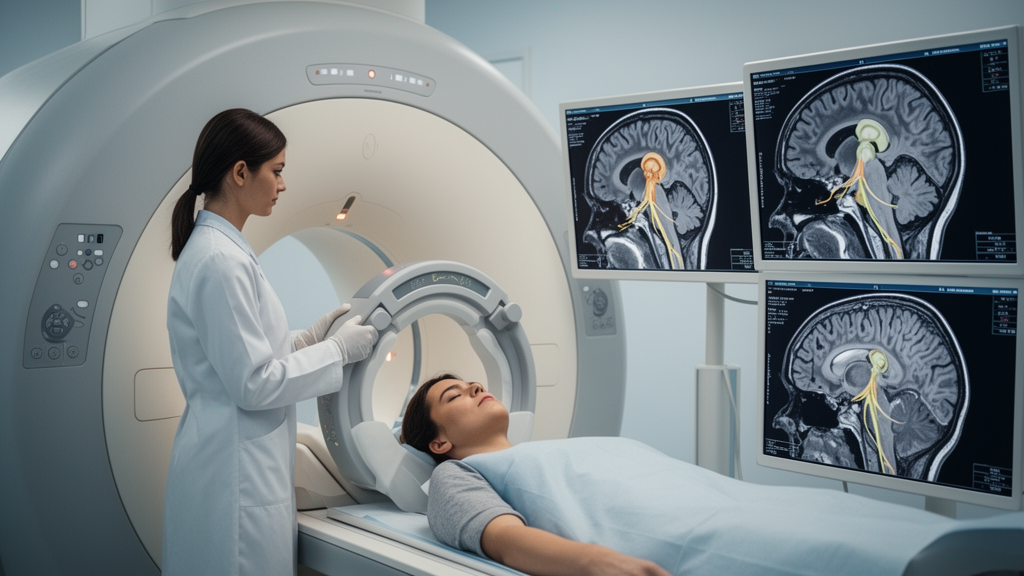
All scans were acquired with a 3.0-T Philips Ingenia scanner and a 16-channel head coil. The investigators used high-resolution sagittal three-dimensional T1-weighted images, acquired with a fast field echo sequence at approximately 1.0-mm isotropic resolution and reconstructed at approximately 0.5-mm resolution. DeepCERES version 1.1, running on the volBrain platform, processed the images through denoising, registration to the Montreal Neurological Institute 152 standard space, bias-field correction, intensity normalization and intracranial-cavity segmentation. The pipeline generated synthetic T2-weighted images and subject-specific multi-atlas priors before an ensemble model delineated cerebellar gray matter, white matter and bilateral lobules. The preoperative and postoperative scans were processed independently rather than through a joint longitudinal segmentation pipeline.
For each participant, the researchers extracted absolute volume and MRI-derived mean cortical thickness from 12 bilateral cerebellar regions: Lobules I–II, III, IV, V, VI, Crus I, Crus II, VIIb, VIIIa, VIIIb, IX and X. Bilateral volume was the sum of the left and right absolute volumes in cubic centimetres, while bilateral thickness was the arithmetic mean of the two sides in millimetres. The measurements also included intracranial volume and the absolute volumes of the whole cerebellum, cerebellar gray matter and cerebellar white matter. DeepCERES thickness is a macroscopic estimate based on MRI contrast and segmentation boundaries; it is not a histological measurement and cannot establish changes in cells or tissue layers.
The main cross-sectional models included diagnostic group, linear and quadratic age terms and sex, while volume models also included intracranial volume. The investigators treated regional volume and regional thickness as separate families of 12 tests and applied the Benjamini–Hochberg procedure to control the false discovery rate within each family. The adjusted thickness difference was 0.104 millimetres for Lobule IV, with a 95% confidence interval of 0.040 to 0.168 and a corrected q value of 0.020. Lobule VI was 0.071 millimetres thicker, with a 95% confidence interval of 0.021 to 0.121 and q equal to 0.036. Neither the remaining lobules nor the regional, whole-cerebellum, cerebellar gray-matter or cerebellar white-matter volume measures showed a significant group difference after correction. The confidence intervals were not themselves adjusted for multiple comparisons.
Sensitivity analysis indicated that the Lobule IV result was the more dependable of the two cross-sectional findings. It remained significant across three approaches to age, with adjusted differences of 0.104 to 0.105 millimetres, and it also remained significant when intracranial volume or automated image-quality scores were added to the model. Lobule VI was more sensitive to analytical choices: its adjusted difference was 0.057 millimetres under a linear age model and did not meet the corrected significance threshold, although a restricted cubic spline model produced a similar estimate of 0.073 millimetres that did pass the threshold. Neither result reversed direction in leave-one-out analyses, and bootstrap confidence intervals excluded zero. Within the trigeminal neuralgia group, however, Lobule VI thickness was positively correlated with automated quality scores, indicating that image quality and the measured structure were not entirely independent.
Disease duration, pain side and imaging-based Sindou grade were not significantly associated with regional volume or thickness after correction for multiple testing. Exploratory analyses also found no pain-related cerebellar asymmetry that passed the corrected threshold. The longitudinal group contained 33 patients who underwent microvascular decompression and 16 who underwent a percutaneous intervention. Clinical follow-up information was incomplete: among 26 patients with available immediate postoperative pain records, 21 had a recorded 100% reduction in pain. The first follow-up was reported for those 26 patients, and second telephone follow-up pain scores were available for 24. The study did not test whether changes in cerebellar morphology were associated with pain improvement, limiting interpretation of the imaging changes as clinically meaningful.
Within the 49 paired scans, postoperative volume increased significantly in Lobules IV, V, VI, VIIb, VIIIa and VIIIb. Mean absolute increases ranged from 0.044 to 0.103 cubic centimetres, while the mean of individual percentage changes ranged from 0.86% to 1.69%. Thickness increased in Lobules IV, V and VI: Lobule IV rose by 0.039 millimetres, Lobule V by 0.066 millimetres and Lobule VI by 0.037 millimetres, with corrected q values of 0.003, 0.002 and 0.003. The corresponding mean individual percentage changes were 1.82%, 3.00% and 1.38%. Supportive analyses also showed increases of 0.594 cubic centimetres in whole-cerebellum volume and 0.538 cubic centimetres in cerebellar gray-matter volume, although white-matter volume did not change significantly after correction.
These small postoperative signals were generally supported by alternative statistical tests, but their stability varied among regions. Nonparametric paired tests and bootstrap resampling supported all nine primary volume and thickness increases, and none reversed direction when individual patients were omitted. Models that simultaneously accounted for changes in intracranial volume and automated quality scores retained significant estimates for volume in Lobules IV, V and VIIIb and thickness in Lobules IV–VI. The volume increases in Lobules VI, VIIb and VIIIa did not pass the corrected threshold in those models, and the leave-one-out results for VIIb and VIIIa were less stable. The direction of the findings also matters: Lobules IV and VI were already thicker between groups, so their postoperative increases cannot be interpreted as normalization toward healthy-control levels.
Several limitations constrain what can be concluded. The cross-sectional design cannot establish whether cerebellar differences preceded or resulted from trigeminal neuralgia, and the longitudinal cohort lacked repeated scans in controls or matched test–retest measurements from the same software version and scanning conditions. Earlier repeat-scan work cited by the authors reported coefficients of variation ranging from 0.70% to 2.95% for unilateral lobular volume and from 0.57% to 1.60% for unilateral thickness in regions corresponding to some findings here, but coefficients of variation and mean changes measure different aspects and cannot show whether the postoperative shifts exceeded measurement error. Most patients also took medication, usually including carbamazepine, with insufficient information on dose, treatment duration and postoperative changes to separate medication effects from disease effects. Visual quality review was not blinded to diagnosis or scan time, and the two procedures were combined despite their potential differences. The results therefore identify MRI-derived morphometric patterns for further study, not a cellular mechanism, causal treatment effect or demonstrated link between postoperative brain change and pain relief.
Subject of Research: MRI-based cerebellar lobular morphometry in patients with trigeminal neuralgia
Article Title: Cerebellar lobular morphometry in trigeminal neuralgia: cross-sectional and exploratory postoperative analyses
Article References: Li, H., Chen, J., Wang, X., She, J., Gao, W., & Wu, T. (2026). Cerebellar lobular morphometry in trigeminal neuralgia: cross-sectional and exploratory postoperative analyses. Frontiers in Neurology, 17, Article 1964745. https://doi.org/10.3389/fneur.2026.1964745
Image Credits: AI Generated
DOI: 10.3389/fneur.2026.1964745
Keywords: trigeminal neuralgia, cerebellar morphology, cortical thickness, MRI, DeepCERES, postoperative imaging, morphometry, cerebellar lobules, longitudinal analysis, neuroimaging, Cerebellar, lobular
News Source: Ophelia Keating. (October 22, 2026). MRI links trigeminal neuralgia to cerebellar thickness and postoperative changes. Scienmag.