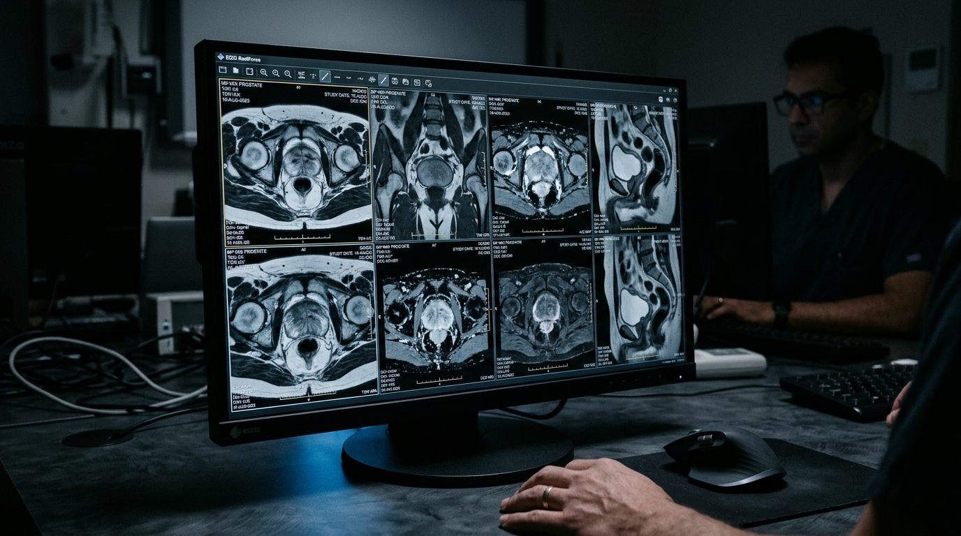
Researchers at two medical centers developed and externally validated a lesion-independent biparametric MRI framework designed to identify pathologically high-risk prostate cancer before treatment. The study, published in Cancer Imaging, utilized whole-gland habitat analysis to characterize intraprostatic heterogeneity without requiring the manual delineation of specific index lesions. The team assessed the predictive value of this habitat framework and evaluated whether adding a prespecified capsule-proximal periprostatic adipose tissue extension could further improve the identification of high-risk disease in patients with pathologically confirmed prostate cancer.
The cohort was divided into two distinct groups for the study design: 241 patients were used for the development of the predictive models, while an independent group of 60 patients served as the external validation cohort. Pathologically high-risk prostate cancer was strictly defined for the purposes of this study as having a Gleason score of 4+3 or higher, or an International Society of Urological Pathology grade group of 3 or higher.
The methodological core of the study involved generating patient-specific whole-gland habitats using joint k-means clustering. The researchers analyzed T2-weighted images and apparent diffusion coefficient maps simultaneously to create two distinct habitat clusters, designated as K = 2. Habitat-derived radiomic signatures were extracted from these clusters and combined with a radiomics signature derived from the periprostatic adipose tissue within 0 to 3 mm of the prostate capsule. These imaging features were then fused with log-transformed prostate-specific antigen density in score-level fusion models to create a comprehensive predictive score for each patient.
Performance evaluation for the development cohort was conducted using stratified five-fold cross-validation, with out-of-fold predictions used to estimate the model’s accuracy.
In the external validation cohort, conventional whole-gland radiomics combined with log-transformed prostate-specific antigen density achieved an area under the receiver operating characteristic curve of 0.791, with a 95% confidence interval ranging from 0.664 to 0.899. The standalone radiomics signature derived from the periprostatic adipose tissue achieved a lower AUC of 0.702, with a 95% confidence interval from 0.540 to 0.845. The H1-derived gland fusion model demonstrated superior performance, achieving an AUC of 0.836 with a 95% confidence interval of 0.715 to 0.936. When the H1-derived signature was extended to include the periprostatic adipose tissue features, the AUC was 0.840, with a 95% confidence interval of 0.710 to 0.941.
The incremental value of adding the periprostatic adipose tissue features to the H1-derived gland fusion model was found to be minimal. The change in AUC was only 0.003, with a 95% confidence interval ranging from -0.046 to 0.048, and a bootstrap p-value of 0.881, indicating that this difference was not statistically significant. Furthermore, the H1-derived gland fusion model exhibited better calibration and predictive accuracy metrics compared to the extended model. It had a lower Brier score of 0.146 compared to 0.175 for the extended model, and a lower expected calibration error of 0.153 compared to 0.219.
The authors conclude that in this small independent external validation cohort of 60 patients, the lesion-independent whole-gland habitat framework showed promising discrimination between pathologically high-risk and lower-risk prostate cancer. The addition of capsule-proximal periprostatic adipose tissue features produced only a negligible increase in the area under the curve and did not reduce prediction or calibration error or increase the net benefit. However, the authors emphasize that larger multicenter studies are necessary to confirm these external findings and to determine whether the periprostatic adipose tissue provides any meaningful incremental predictive value beyond the H1-derived gland fusion model.
Subject of Research: Biparametric MRI habitat analysis for identifying high-risk prostate cancer
Article Title: Biparametric MRI–based whole-gland habitat analysis for pretreatment identification of pathologically high-risk prostate cancer: a two-center study with external validation
Article References: Huang, J., Gong, J., Zhang, Y., Yang, O., Li, J., Chen, J., & Huang, G. (2026). Biparametric MRI–based whole-gland habitat analysis for pretreatment identification of pathologically high-risk prostate cancer: a two-center study with external validation. Cancer Imaging. https://doi.org/10.1186/s40644-026-01139-0
Image Credits: AI Generated
DOI: 10.1186/s40644-026-01139-0
Keywords: prostate cancer, MRI, habitat analysis, radiomics, diagnostic imaging, oncology, prostate-specific antigen, medical imaging, cancer risk, external validation, Biparametric, based
News Source: Nathaniel Bowman. (November 7, 2026). MRI Habitat Analysis Identifies High-Risk Prostate Cancer. Scienmag.