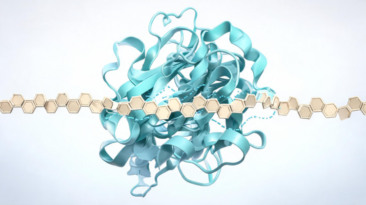
Deep beneath the soil, a microscopic war is being waged between fungi, and one of the most important weapons in that conflict has now been mapped in unprecedented detail. Researchers have turned to molecular modeling and simulation to dissect the chitinase enzyme produced by Paraphaeosphaeria minitans, a mycoparasitic fungus long prized as a biological control agent against the devastating plant pathogen Sclerotinia. By building a three-dimensional model of the enzyme, docking it with its substrate chitotriose, and running extensive molecular dynamics simulations, the team has revealed the structural choreography that allows this enzyme to bind and degrade chitin, the tough polymer that forms the skeletons of insects and the cell walls of fungi. The findings, published in Discover Green Chemistry, offer a molecular blueprint that could guide the engineering of more potent biopesticides and industrial enzymes.
The significance of the work begins with the target itself. Chitin, composed of repeating units of N-acetyl-D-glucosamine linked by beta-1,4-glycosidic bonds, is the second most abundant biopolymer in nature, found in insect cuticles, fungal cell walls, and even some plants. Chitinases, the enzymes that cleave these bonds, belong to glycoside hydrolase families GH18 and GH19, and those in the GH18 family fold into a characteristic alpha/beta barrel domain that carries out catalysis. Fungal chitinases are of particular interest because they can be deployed against agricultural pests and pathogens, dissolving the protective armor of organisms such as Colletotrichum gloeosporioides, Rhizoctonia solani, Fusarium graminearum, and Fusarium oxysporum. Yet despite their promise, the structural and functional properties of fungal chitinases remain poorly charted, largely because experimental techniques such as X-ray crystallography are costly and technically demanding.
Paraphaeosphaeria minitans, formerly known as Coniothyrium minitans, is a mycoparasite that attacks Sclerotinia species, a group of fungal pathogens that infect more than 400 plant species, including soybeans, sunflowers, canola, and beans. The economic stakes are enormous: outbreaks of Sclerotinia stem rot caused losses of roughly 227 million dollars in 1997, 344 million dollars in 2004, and 560 million dollars in 2009. P. minitans invades the pathogen’s resilient sclerotia, and its chitinase is central to this parasitic process, helping the mycoparasite penetrate and dismantle the fungal cell walls of its host. Understanding how this enzyme works at the atomic level is therefore not just an academic exercise but a step toward more effective and sustainable crop protection.
To build their model, the researchers retrieved the protein sequence of the enzyme, designated PmChi, from the UniProt database and employed homology modeling powered by deep learning through the RaptorX platform, followed by refinement with DeepRefiner. The resulting structure was validated with a battery of computational tools, including Procheck, ProSA, Verify 3D, and ERRAT. The model showed a Z-score of -7.21, indicating that most residues sit in energetically favorable states, and a QMEAN score of 0.65, confirming reliability. Ramachandran analysis placed 79.6 percent of residues in the most favored regions, and the ERRAT quality score reached 90.824, all pointing to a structurally sound model suitable for downstream analysis.
The physicochemical portrait of PmChi proved equally informative. The enzyme has a molecular weight of 47 kilodaltons and a theoretical isoelectric point of 4.96, reflecting an abundance of acidic amino acids that also favor protein solubility. Its instability index of 32.60 falls below the threshold of 40, predicting stability in vivo, while an aliphatic index of 74.94 suggests thermostability. Secondary structure analysis revealed that random coils dominate at 51.24 percent, followed by alpha helices at 28.44 percent and extended strands at 14.22 percent. Two N-glycosylation sites were identified, which likely assist secretion, folding, and stability. Sequence analysis confirmed that PmChi belongs to glycosyl hydrolase family 18, subfamily II, spanning residues 52 to 416, and contains a chitinase insertion domain composed of antiparallel beta strands and an alpha helix nested within the TIM barrel.
At the heart of the enzyme lies a conserved catalytic motif. GH18 chitinases typically carry the sequence DxxDxDxE, and PmChi carries FDGLDIDWE at positions 178 to 186. Within this motif, the glutamic acid acts as the catalytic proton donor, while the aspartic acid stabilizes the protein by accommodating substrate distortion during the reaction. Docking studies with AutoDock 4.2 identified the key residues involved in substrate recognition and catalysis: Trp146, Asp184, Glu186, Tyr187, Pro228, Met252, Tyr254, Asp255, Trp393, Arg310, and Glu395. The calculated Gibbs free energy for binding chitotriose was -6.62 kilocalories per mole, indicating a spontaneous and stable interaction, with a ligand binding efficiency of -0.196 kilocalories per mole and a pKd of 4.94, suggesting moderate affinity with room for optimization through protein engineering.
The molecular dynamics simulations, run for 200 nanoseconds with GROMACS using the CHARMM27 force field, delivered the study’s most striking dynamic insights. The root mean square deviation of the free enzyme stabilized at approximately 1 nanometer, but the PmChi-chitotriose complex settled at around 0.5 nanometer and showed none of the early fluctuations seen in the unbound protein between 0 and 80 nanoseconds. This enhanced stability reflects the anchoring effect of chitotriose binding, which locks the active site into a catalytically competent conformation through hydrogen bonds, hydrophobic contacts, and van der Waals forces. Root mean square fluctuation analysis reinforced the picture: free PmChi displayed greater residue flexibility, while the ligand-bound complex was markedly more rigid, underscoring how substrate binding restrains protein motion and preserves the structural integrity of the active site during catalysis.
Hydrogen bond analysis revealed that the enzyme-carbohydrate interaction was maintained by four to ten hydrogen bonds throughout the simulation, with Tyr254 and Trp185 emerging as the principal residues forming these contacts. Solvent accessibility calculations showed that the enzyme cavity, initially highly exposed to solvent, became progressively less accessible over the 200-nanosecond trajectory, suggesting that chitotriose binding induces a more closed and compact conformation that stabilizes the enzyme-substrate complex. Binding free energy calculations using both MM/GBSA and MM/PBSA methods decomposed the interaction residue by residue. In the MM/GBSA analysis, Glu186 contributed approximately -7.0 kilocalories per mole, the largest single contribution, followed by Trp146 and Asp255 at around -4.0 each, while Arg310 was the only residue contributing unfavorably. The Poisson-Boltzmann-based MM/PBSA approach proved more accurate for analyzing enzyme-carbohydrate interactions, consistent with its theoretical rigor.
Beyond the active site, the simulations illuminated the broader interaction network that keeps PmChi stable. Hydrophobic bonds increased from 702 before the simulation to 827 after it, revealing a non-polar protein surface primed for intermolecular interactions even as the active site itself is rich in catalytic aspartate and glutamate residues. The enzyme also maintained 16 pi-pi stacking interactions before the simulation and 14 after, interactions known to support protein stability, carbohydrate recognition, catalysis, and self-assembly. Salt bridge analysis identified six such interactions, dominated by Lys/Asp pairs at 59 percent, with Arg/Asp pairs accounting for 22 percent, all shorter than 4.0 angstroms. These non-covalent interactions collectively explain how the enzyme withstands the conformational stresses of substrate binding and hydrolysis in an aqueous environment.
The study’s authors note that extending simulations beyond 500 nanoseconds will help delineate the long-term structural stability and interaction dynamics of the PmChi-chitotriose complex. Even now, however, the work delivers a platform for future research and applications. By pinpointing the residues that govern substrate binding and catalysis, and by demonstrating that ligand binding stabilizes rather than destabilizes the enzyme, the study provides a rational starting point for protein engineering aimed at boosting catalytic efficiency or tailoring substrate specificity. In a world seeking alternatives to synthetic pesticides, understanding the molecular mechanics of a fungus that parasitizes crop pathogens is a powerful step toward greener agriculture, and this computational portrait of PmChi brings that goal measurably closer.
The broader context of biocontrol helps explain why this enzyme matters. Microbial inoculants such as P. minitans suppress plant disease through several complementary mechanisms, including the secretion of cell-wall-degrading enzymes, the release of siderophores that sequester iron from the rhizosphere, competition for space and nutrients, and the induction of systemic resistance within the plant itself. Chitinase production sits squarely within this enzymatic arsenal, physically dissolving the chitin-rich walls of target fungi and enabling the mycoparasite to breach its host.
The products of chitin hydrolysis also carry commercial weight. Chitooligomers liberated by chitinases are finding uses across agriculture, food processing, and biomedicine, which means an enzyme optimized for efficient chitin cleavage could serve dual purposes: strengthening biological control in the field and supplying valuable oligosaccharides for downstream applications. This dual utility aligns the work with green chemistry goals, replacing synthetic pesticides and harsh chemical depolymerization routes with enzymatic alternatives.
The computational strategy itself deserves note. Homology modeling guided by deep learning, combined with docking and extended molecular dynamics, offers a route to structural insight when experimental crystallography is impractical, and the validation metrics reported here suggest such models can be reliable enough to nominate specific residues for mutagenesis. The finding that MM/PBSA outperformed MM/GBSA for this enzyme-carbohydrate pair adds a practical benchmark for future simulation studies of glycoside hydrolases.
Finally, the moderate binding affinity observed with chitotriose hints at natural substrate tuning. Because chitin in fungal walls is polymeric, the enzyme likely engages longer chains in vivo, and the residues identified here provide concrete targets for engineering variants with enhanced processivity, thermostability, or antifungal potency tailored to specific crop pathogens.
Subject of Research: Molecular dynamics analysis of the chitinase enzyme from the mycoparasitic biocontrol fungus Paraphaeosphaeria minitans and its interaction with chitotriose
Article Title: Unveiling the interaction between fungal biocontrol agent Paraphaeosphaeria minitans chitinase and chitotriose through molecular dynamics study
Article References: Jana, U. K., & Kango, N. (2026). Unveiling the interaction between fungal biocontrol agent Paraphaeosphaeria minitans chitinase and chitotriose through molecular dynamics study. Discover Green Chemistry, 1(1), Article 30. https://doi.org/10.1007/s44509-026-00029-9
Image Credits: AI Generated
DOI: 10.1007/s44509-026-00029-9
Keywords: chitinase, Paraphaeosphaeria minitans, molecular dynamics, chitotriose, biocontrol, GH18, homology modeling, molecular docking, MM/PBSA, Sclerotinia, chitin, protein engineering
Cite Scienmag News
APA MLA Chicago
Bethany Barker. (September 10, 2026). Molecular Dynamics Reveal How a Fungal Biocontrol Enzyme Grips Chitin. Scienmag. https://scienmag.com/molecular-dynamics-reveal-how-a-fungal-biocontrol-enzyme-grips-chitin/
Bethany Barker. “Molecular Dynamics Reveal How a Fungal Biocontrol Enzyme Grips Chitin.” Scienmag, 10 September 2026, https://scienmag.com/molecular-dynamics-reveal-how-a-fungal-biocontrol-enzyme-grips-chitin/. Accessed 10 September 2026.
Bethany Barker. “Molecular Dynamics Reveal How a Fungal Biocontrol Enzyme Grips Chitin.” Scienmag. September 10, 2026. https://scienmag.com/molecular-dynamics-reveal-how-a-fungal-biocontrol-enzyme-grips-chitin/
Copy citation Download RIS
Tags: biocontrolchitinchitin degradation mechanismchitinasechitinase engineering for industrial applicationschitotriosedesign of biopesticides targeting chitinenzyme-substrate docking of chitotriosefungal biocontrol agents against plant pathogensfungal biocontrol enzymefungal cell wall biopolymer breakdownGH18homology modelingMM/PBSAmolecular blueprint for enzyme enhancementmolecular dockingmolecular dynamicsmolecular dynamics simulation of chitinasemolecular modeling of chitinaseParaphaeosphaeria minitansParaphaeosphaeria minitans enzyme structureProtein EngineeringSclerotiniastructural analysis of glycoside hydrolases