In a groundbreaking advance at the crossroads of biophysics and photonics, researchers at the University of Cologne have engineered ultra-small, flexible microlasers capable of measuring mechanical forces inside living cells. These novel microlasers, which are only about 20 microns in diameter—roughly the width of a human hair—exploit whispering gallery mode resonance to trap and amplify light within a tiny elastomer bead doped with fluorescent dye. This extraordinary development opens new frontier avenues for probing biomechanical properties at the cellular level, which has profound implications for understanding processes such as embryonic development, tissue morphogenesis, and cancer metastasis.
Mechanical forces operating within and between cells orchestrate a myriad of biological functions and pathologies. Cells constantly generate and experience forces as they adhere, migrate, divide, and remodel their surrounding environment. In cancer, for instance, malignant cells invade neighboring tissues by physically squeezing through the dense cellular matrix. Until now, it has been challenging to directly quantify these pico- to nanonewton-scale forces within live cell environments due to limitations in existing techniques. The implementation of these microlasers offers a new paradigm—using optical signals rather than conventional mechanical or fluorescent markers—to measure forces with high spatial and temporal precision.
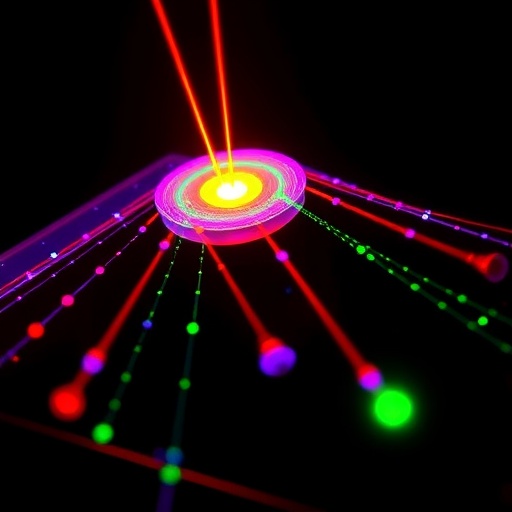
The microlasers capitalize on whispering gallery mode phenomena, where light circulates along the inner circumference of a spherical cavity, effectively trapped by continuous total internal reflection. This circulating light amplifies coherently to form a laser when the cavity incorporates a gain medium, in this case, fluorescent dye integrated into a flexible elastomer bead. The key innovation lies in balancing the mechanical softness of the material—allowing deformation under forces exerted by cells—with a sufficiently high refractive index to sustain laser action. Achieving this required identifying a commercially available elastomer optimized for both flexibility and optical performance, a challenge given that materials with high refractive indices tend to be rigid.
When external forces are applied to these microbeads, the shape and internal strain of the bead change, which in turn shifts the spectral properties of the laser emission. By monitoring these shifts, researchers can quantitatively correlate changes in laser wavelength and mode width with precise force magnitudes up to around 50 nanonewtons. Such sensitivity neatly matches the biomechanical range pertinent to intercellular forces, enabling direct biophysical insights without perturbing the natural cellular milieu. Moreover, this laser-based sensing does not require direct imaging, an advantage in dense tissue contexts where optical access is limited or where fluorescence-based probes suffer from photobleaching and background noise.
Unlike prior efforts that relied on liquid droplet lasers, which are often too soft and unstable for biological force measurements, these elastomer-based microlasers exhibit robust mechanical stability with Young’s modulus values comparable to living cells. This mechanical compatibility facilitates cellular internalization—the beads can be readily taken up by cells via endocytosis without disrupting normal physiology. Long-term stability in cell culture conditions was verified, confirming that the microlasers maintain lasing performance over several days, a vital attribute for longitudinal force monitoring in dynamic biological processes.
The research team employed an atomic force microscope as a calibration tool to mechanically manipulate single microbeads and simultaneously record their laser emission spectra. This meticulous calibration enabled them to construct precise force-to-optical signal maps. The microlasers demonstrated multimodal lasing thresholds as low as 2 nanojoules, underscoring their high optical efficiency. Notably, the spectral mode widths broadened proportionally with applied force, providing an additional parameter for refining force quantification.
These versatile microlasers herald a new class of biosensors that can be tailored to varying biomechanical sensitivities by adjusting the elastomer’s stiffness and dye concentration. In future iterations, the team envisions applications beyond cellular biomechanics, including probing contractile forces in muscle and heart tissues. The ability to measure forces deep within tissues, where conventional imaging modalities are impractical, could revolutionize mechanobiology and biomedical diagnostics by delivering spatially resolved force maps in vivo.
Apart from the immediate technological breakthrough, this innovation also underscores the vital synergy between materials science and biophotonics. Designing a soft yet optically capable lasing medium required navigating trade-offs between mechanical deformability and optical confinement, a balance rarely achieved in prior microlaser work. The highly reproducible fabrication process, enhanced by size uniformity and chemical stability, ensures that these microlasers can be mass-produced and tailored for diverse biological investigations.
Looking ahead, ongoing research is focused on enhancing the long-term photostability and reproducibility of the microbeads, crucial for clinical and experimental deployment. By standardizing fabrication parameters and exploring alternative dye chemistries, the researchers aim to mitigate variability and extend the operational lifespan of these biosensors under physiological conditions. Such improvements will empower continuous monitoring of biomechanical fluctuations during complex biological events like tissue regeneration and cancer invasion.
In essence, this pioneering work paves the way for integrating optical microlasers as minimally invasive biomechanical probes within living systems. By transforming mechanical stress into easily measurable optical signals, these whispering gallery mode microbeads could unlock unprecedented understanding of the physical forces that shape life at the microscopic scale. As mechanobiology continues to emerge as a crucial frontier in biomedical science, innovations like these microlasers will be indispensable tools in the quest to decipher the language of cellular forces.
Subject of Research: Biomechanical force sensing in living cells using elastomer-based whispering gallery mode microlasers.
Article Title: Elastomer-based whispering gallery mode microlasers with low Young’s modulus for biosensing applications.
News Publication Date: Information not provided.
Web References: https://doi.org/10.1364/OME.600106
References: M. Bayrak, D. Ripp, J. S. Hill, M. Schubert. “Elastomer-based whispering gallery mode microlasers with low Young’s modulus for biosensing applications,” Optical Materials Express 16, 1440-1453 (2026).
Image Credits: Marcel Schubert, University of Cologne
Keywords: Microlasers, whispering gallery mode, elastomer, biosensing, cellular force measurement, biomechanical sensing, photonics, fluorescent dye, nanonewton forces, cell mechanics, mechanobiology.
Tags: biomechanical force measurement inside living cellscellular biomechanics and tissue morphogenesisfluorescent dye-doped elastomer beadshigh precision biomechanical force sensorsminiature flexible microlasers for cellular force sensingnanonewton-scale force quantification in cancer metastasisnon-invasive cellular force measurement methodsoptical force sensing techniques in live cellsprobing mechanical forces in embryonic developmentultra-small microlasers for biophysics researchUniversitywhispering gallery mode resonance in biophotonics


