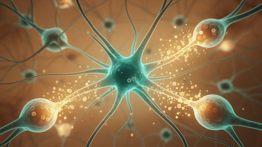
Deep inside the developing brain, the resident immune cells known as microglia are quietly running one of biology’s most consequential construction projects. First described in 1919 by Spanish neuroscientist Pio del Rio-Hortega as the third glial cell, these mesoderm-derived scavengers were long dismissed as dormant sentinels that only sprang into action after injury or infection. Intravital imaging in live animals shattered that view, revealing that microglia in their so-called resting state are extraordinarily dynamic, constantly surveying their surroundings with motile processes. Researchers now prefer the term homeostatic microglia, and decades of work have established these cells as indispensable architects of brain wiring, contributing to neurogenesis, developmental neuronal apoptosis, synaptic pruning, and the secretion of neurotrophic factors. When microglial programs falter, the consequences reach from neurodevelopmental disorders to neurodegeneration, and genome-wide association studies have repeatedly implicated microglial immune genes in psychiatric illness, autism, and schizophrenia.
What makes microglia such powerful modulators of the nervous system is the vast signaling repertoire they deploy. They secrete and respond to cytokines, chemokines, complement proteins, membrane-associated recognition molecules, and reactive oxygen species, each class acting at specific stages of neural development from precursor cell proliferation to the final refinement of synaptic connections. While microglia supply the bulk of immune surveillance in the developing brain, they are not alone. Maternally derived microchimeric microglia help colonize the progeny’s brain and stabilize the microglial population, yolk sac-derived border-associated macrophages populate the brain during development, and adaptive immune cells such as B-1a lymphocytes and CD4-positive T cells promote oligodendrogenesis and microglial maturation respectively. Human evidence is now accumulating through brain organoids, xenotransplanted human microglia, single-cell RNA sequencing of fetal microglia, and induced pluripotent stem cell-derived microglia in culture.
Among the chemokines, fractalkine has emerged as a master regulator of the neuron-microglia dialogue. This membrane-bound protein, which can be cleaved into a soluble form, binds exclusively to the CX3CR1 receptor on microglia, and neurons secrete it to attract microglial processes while maintaining brain homeostasis and neuronal viability. Correlative human data are striking: CX3CR1 expression is downregulated in schizophrenia patients, fractalkine levels are lower in autism spectrum disorder cohorts, and the Ala55Thr variant of CX3CR1 is genetically associated with both conditions. In rodents, maternal immune activation triggered by poly(I:C) or lipopolysaccharide decreases microglial CX3CR1 expression alongside working memory and social behavior impairments. Knockout experiments reveal the developmental stakes. CX3CR1-deficient mice show reduced microglial colonization of the hippocampus and somatosensory cortex, more apoptotic neurons in the motor cortex, excess PSD95 puncta, dendritic spines, and immature synapses at postnatal day 15, and delayed maturation of NMDA receptor kinetics at thalamocortical synapses. Fractalkine knockout mice display long-term memory and spatial learning deficits tied to impaired hippocampal neurogenesis and long-term potentiation, deficits that exogenous soluble fractalkine can rescue.
Interleukin-17A tells a more cautionary tale about dosage. Expression of this cytokine is very low in the developing brain, yet its receptor is highly expressed in microglia, and its structural similarity to nerve growth factor has led researchers to postulate neurotrophic properties. Human studies link elevated serum IL-17A to autism in children, although one study reports a significant decrease, and levels are reduced in children born after maternal Zika virus infection who exhibit microcephaly. Applying IL-17A to human iPSC-derived neural progenitor cells increases synaptic and neuronal polarity proteins, while neonatal IL-17A knockout mice show decreased synaptophysin and PSD95. Conversely, gestational increases driven by prenatal stress, SARS-CoV-2 spike protein exposure, poly(I:C), or recombinant IL-17 protein injection correlate with altered microglial morphology, cortical anomalies, and behavioral changes in offspring. IL-17A also amplifies the inflammatory response by promoting production of IL-1β, TNF-α, and IL-6, suggesting that both excess and deficiency of this single cytokine can derail developmental programs.
Interleukin-6 and interleukin-1β illustrate how tightly the immune system must calibrate its signals for the brain to wire correctly. Microglia express IL-6, its receptor, and the signaling subunit glycoprotein 130, positioning them to mediate many of IL-6’s developmental functions, which include inducing early neurogenesis and neuronal differentiation of human neural stem cells, promoting neuron survival in culture, yet significantly reducing adult hippocampal neuronal differentiation through the JAK/STAT3 pathway. In a murine maternal immune activation model, microglia at embryonic day 16.5 adopt an inflammatory phenotype in wild-type but not IL-6 knockout fetuses, while non-microglial fractions from IL-6 knockout mice paradoxically show high basal inflammatory cytokine expression even under saline controls, underscoring the need for precisely controlled levels. IL-1β, produced in the central nervous system by microglia, is expressed at high levels during development and modulates astrocyte progenitor migration through calcium signaling, contributes to neural precursor proliferation and differentiation likely via the Wnt5a/RhoA/JNK pathway, acts synergistically with neurotrophin-3 to stimulate neurite outgrowth, and guides radial migration of cortical neurons, since RNAi inhibition of its receptor decreases migration at postnatal day 5. In zebrafish, both elevated and inhibited IL-1β alter retinal ganglion cell axon arborization, and an IL-1β gene polymorphism is associated with increased autism incidence in humans.
Tumor necrosis factor alpha demonstrates that even a classic pro-inflammatory cytokine can serve constructive ends when levels are closely controlled. High concentrations induce apoptosis of cultured neonatal neural precursor cells, while low concentrations promote proliferation and differentiation. TNF-α acts as a permissive factor for glutamate release in astrocytic gliotransmission, modulates the composition of GABA and glutamate receptor subunits at neuronal membranes, and astrocyte-derived TNF-α drives the homeostatic up-scaling of AMPA receptor-mediated synaptic transmission in response to chronic activity blockade and monocular deprivation during the ocular dominance critical period. In the developing Xenopus tadpole visual system, chronic TNF-α exposure accelerates the unsilencing of immature NMDA receptor-only synapses by trafficking AMPA receptors to retinotectal synapses and expands dendritic arbors of optic tectal neurons. Interferon-γ, meanwhile, negatively regulates the neural stem cell niche, with its inhibitory effect paradoxically stronger in the absence of microglia, hinting that microglia release protective factors, and interferon-γ knockout mice show behavioral impairments and defects in neuronal connectivity. Astrocyte-derived IL-33 recruits microglia to refine synapses: conditional deletion in astrocytes increases excitatory and inhibitory synapse numbers and reduces microglial engulfment of PSD95 puncta, while excess IL-33 reduces VGLUT-1 synapses and boosts engulfment, and loss of the microglial ST2 receptor impairs spine plasticity, newborn neuron integration, and memory.
Beyond soluble messengers, microglia rely on membrane recognition systems to decide which synapses live and which are eaten. Phosphatidylserine, an anionic phospholipid normally confined to the inner leaflet by flippase enzymes, is externalized during injury or apoptosis and serves as a canonical eat-me cue. Activity regulates this process: knockdown of the flippase chaperone CDC50A increases microglial engulfment of presynaptic terminals in the retinogeniculate projection, and postsynaptic CDC50A knockout selectively eliminates inhibitory synapses, an effect prevented by knockout of the microglial phagocytic receptor MERTK. Additional PS receptors shape the outcome. GPR56 knockout mice show reduced microglial engulfment and increased synapse densities in the dorsal lateral geniculate nucleus and hippocampus, TREM2 knockout mice display higher densities of excitatory synaptic markers and increased miniature EPSC frequency, and in embryonic zebrafish, BAI-1 knockdown doubles phagosome formation time while TIM-4 knockout creates unstable phagosomes that lose their cargo. Purinergic P2Y12 receptors, activated by ADP, mobilize microglia toward neuronal activity, form dynamic somatic microglial junctions with pyramidal neuron cell bodies detected as early as embryonic day 15, and their loss produces aberrant cortical layer formation, increased hippocampal excitability, anxiety-like behavior, and mutations in the encoding locus are observed in some epileptic patients.
The complement system provides the molecular tagging machinery for synaptic elimination. In the classical pathway, C1q within the C1 complex recognizes targets, activating the C1r and C1s proteases that cleave C4 and C2 to form the C3 convertase, which cleaves C3 into C3a and the C3b opsonin that signal through C3aR and CR3 on microglia to promote engulfment. C1q expression is punctate and localized to synapses during development, C1q knockout mice show impaired synapse elimination, and C1q has recently been found to bind phosphatidylserine directly, suggesting a synergistic targeting mechanism. C3 knockout increases the neural precursor pool in the subventricular zone, produces cortical migration deficits rescuable by C3a peptide mimics, and in Xenopus, presynaptic C3 overexpression reduces retinal ganglion cell axon arbor length and branch complexity. Genetic risk data converge on this pathway: GWAS strongly links schizophrenia to the MHC locus where copy number variation drives altered C4 expression, C4 knockout mice show reduced eye-specific segregation in the dLGN, and iPSC-derived astrocytes from autism patients show decreased C4 expression, aligning with the hypothesis that schizophrenia involves excessive synaptic elimination while many autism forms involve insufficient pruning.
Just as important as the eat-me signals are the don’t-eat-me signals that spare appropriate contacts. CD47 expressed by developing neurons inhibits phagocytosis through the microglial SIRPα receptor, and mice lacking CD47 show increased engulfment of retinogeniculate terminals and reduced synapse numbers. Xenopus retinal ganglion cells express aRCA3, a putative CD46 homolog whose overexpression reduces axonal engulfment and yields larger, more branched arbors, while the neuronally secreted inhibitors CSMD1 and SRPX2 block complement deposition and C1q activity respectively, with knockout mice showing preferential phagocytosis of synaptosomes or reduced synapse numbers. Even reactive oxygen species, usually cast as damaging radicals, act as physiological second messengers: neural stem cells maintain high intracellular ROS to favor proliferation, hippocampal neurons generate ROS through NADPH oxidases during development, and scavenging superoxide prevents NMDA receptor-mediated long-term potentiation. Caspase-3, long recognized as the point of no return for apoptosis, tags presynaptic sites with phosphatidylserine and C1q during activity-dependent refinement, and Casp3 knockout mice fail to prune functional inputs in the dLGN. Together, these findings reveal the innate immune system as a fundamental regulator of circuit formation, and elucidating its precise pathways promises new therapeutic targets for neurodevelopmental and psychiatric disease.
Subject of Research: Neuroimmunology
Article Title: Microglia and immune signaling as architects of the brain
Article References: Kim, A., Solek, C. M., & Ruthazer, E. S. (2026). Microglia and immune signaling as architects of the brain. Frontiers in Cellular Neuroscience, 20, Article 1936860. https://doi.org/10.3389/fncel.2026.1936860
Image Credits: AI Generated
DOI: 10.3389/fncel.2026.1936860
Keywords: microglia, neurodevelopment, synaptic pruning, complement system, cytokines, fractalkine, neuroimmunology, brain development, phagocytosis, neuroscience, immune, signaling
News Source: Cassandra Pierce. (October 25, 2026). Microglia and immune signaling as architects of the brain. Scienmag.