The malaria parasite Plasmodium vivax may accumulate in the spleen soon after infection to a greater extent than its better-known relative P. falciparum, according to new research published by John Woodford of the University of Queensland, Brisbane, Australia and colleagues in the open access journal PLOS Medicine.
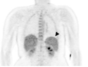
Managing and treating P. vivax and P. falciparum infections calls for investigation of their different pathways of infection, and our limited understanding of disease pathology has generally relied on indirect and imprecise approaches. Woodford and colleagues studied 7 healthy participants who were infected under controlled conditions with either P. vivax or P. falciparum. They underwent a Positron Emission Tomography (PET) scan and Magnetic Resonance Imaging (MRI) 7 days before infection and again 7 to 11 days afterwards, before receiving antimalarial treatment.
The team investigated participants’ spleen, liver and bone marrow for changes in form or structure as well as glucose metabolism, which would suggest accumulation of the parasite in individual organs. In the spleen, glucose metabolism increased following infection, and this was more pronounced in participants infected with P. vivax. Neither the liver nor bone marrow were affected at this early stage of infection. Despite the small size of the study, the research shows that imaging in this way can help in understanding how malarial parasites accumulate in specific organs as well as modifying previous thinking about the behavior of P. vivax at the “blood stage” of infection.
Dr. Woodford notes, “Malaria parasites outside of the circulation contribute to disease but are very difficult to study. By performing this novel imaging study in participants undergoing experimental malaria infection, we have been able to look at what is happening inside specific organs during the earliest stages of blood stage infection. This represents further evidence that parasites of the important species Plasmodium vivax have a particular predilection for the spleen, and highlights how controlled infection studies can accommodate unique exploratory objectives that cannot be otherwise studied in humans.”
###
Research Article
Peer reviewed; Observational; Humans
In your coverage please use this URL to provide access to the freely available paper:
http://journals.
Funding: This project was supported by a HIRF Seed Funding Grant, Metro North Hospital and Health Service (J.W) and by the Australian National Health and Medical Research Council (J.S.M #1135955, #1037304, #1132975, and N.M.A #1135820, 1098334). The clinical trials contributing participants were funded by the Australian National Health and Medical Research Council (J.S.M #1132975), the Bill and Melinda Gates Foundation (J.S.M OPP1111147) and the Global Health Innovative Technology Fund. The funders had no role in study design, data collection and analysis, decision to publish, or preparation of the manuscript.
Competing Interests: The authors have declared that no competing interests exist.
Citation: Woodford J, Gillman A, Jenvey P, Roberts J, Woolley S, Barber BE, et al. (2021) Positron emission tomography and magnetic resonance imaging in experimental human malaria to identify organ-specific changes in morphology and glucose metabolism: A prospective cohort study. PLoS Med 18(5): e1003567. https:/
Media Contact
John Woodford
[email protected]
Related Journal Article
http://dx.


