Risky strategy prepares the parasite for the unexpected but could be exploited to fight the deadly disease
Credit: Penn State
The parasite that causes malaria expresses genes that code for the proteins it will need in later life stages, using two separate schemes to prevent these proteins from actually being made until they are needed, according to new research. Having the mRNAs for these genes at the ready is risky: It’s energetically costly, and proteins made prematurely can cause the parasites to become non-infectious. However, this strategy allows the parasite to quickly respond to unpredictable changes as it is transmitted between its mosquito and human hosts. Understanding these “translational repression” schemes may allow researchers to spot their weaknesses, which could be exploited in new strategies for combatting malaria.
The research, by a team of scientists at Penn State, the Institute for Systems Biology in Seattle, Johns Hopkins, and the University of Washington, appears October 31, 2019 in the journal, Nature Communications.
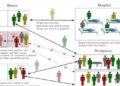
“The malaria parasite has a complex life-cycle in which it is transmitted back-and-forth between its mosquito and human hosts,” said Scott Lindner, assistant professor of biochemistry and molecular biology at Penn State and one of the leaders of the research team. “The parasite can’t predict when these transmissions will happen, so it needs to be able to react quickly to be able to deal with the changes in its environment. We knew how the parasite prepares for the jump from humans into mosquitoes, but until now no one had looked systematically at how the parasite prepared itself for going from mosquitoes into humans.”
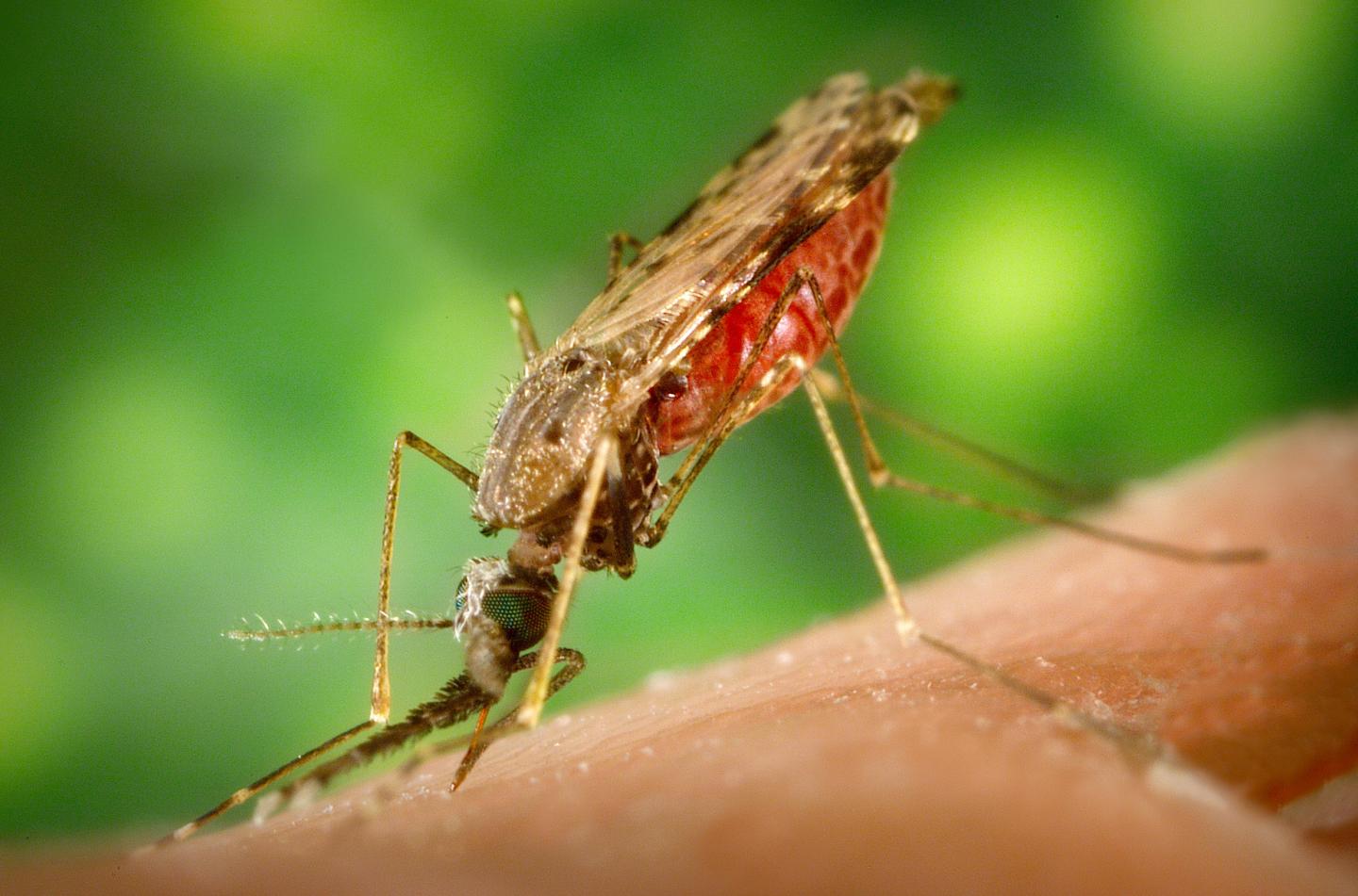
The malaria parasite, which according to the 2018 WHO World Malaria Report affects 200 million people annually, resulting in around 400,000 deaths, enters a mosquito when it takes a blood meal from an infected mammal. Inside the mosquito the male and female parasites fuse and eventually produce an oocyst on the mosquito’s stomach. Inside the oocyst, the parasites undergo further transformation forming thousands of weakly-infectious sporozoites. When an oocyst ruptures, the sporozoites travel through the mosquito’s equivalent of blood and burrow into the mosquito’s salivary glands where they can be transmitted back into a mammal when the mosquito takes its next meal. During this trip from the oocyst and into the salivary gland, the sporozoites become 10,000 times more infectious.
The research team produced and purified large numbers of sporozoites from both the oocyst and salivary-gland stages, then used state-of-the-art RNA sequencing and mass spectrometry-based proteomics to identify essentially all of the mRNAs and proteins that were present in each stage. They did this in both Plasmodium yoelii–a malaria parasite that infects mice, which is easier to handle in the laboratory–and Plasmodium falciparum–a human-infectious parasite that causes most of the documented deaths associated with malaria.
“P. yoelii is often preferred in laboratory studies because we can easily track it through its entire life cycle, but it could have important differences to P. falciparum,” said Lindner. “By studying both, we can determine how conserved these processes are and whether there are specific mRNAs or proteins that behave similarly across the parasite species that infect different hosts.”
Through this study, the researchers demonstrated that, using a scheme similar to the parasites that are transmitted from humans to mosquitoes, the sporozoites produce mRNAs for genes that they will need in the next stage of their life cycle, but then actively repress their translation into proteins. As many as three-quarters of the most abundantly produced mRNAs are translationally repressed in this way.
“Excitingly, we identified two separate translational repression programs that operate simultaneously, independently, and upon different mRNAs,” said Lindner. “The first program represses mRNA produced in oocyst sporozoites, which code for proteins that are made at some point during the parasite’s trip from the mosquito’s midgut into the salivary glands. The second program represses mRNA that are produced throughout both sporozoite stages but doesn’t allow the production of the encoded proteins until the parasite is transmitted into its mammalian host.”
“We are now trying to find how these translational repression programs are controlled in the parasite,” said Lindner, “and if there are weaknesses that we can exploit in this risky strategy that we can use to push the parasite off the edge with new therapeutics.”
###
In addition to Lindner, the research team includes Michael P. Walker, Erin N. Vrana, Kevin J. Hart, and Allen M. Minns at Penn State; Kristian E. Swearingen and Robert L. Moritz at the Institute for Systems Biology in Seattle, WA; Melanie J. Shears and Photini Sinnis at the Johns Hopkins School of Public Health; and Stefan H.I. Kappe at the University of Washington. The research was funded by Penn State, the U.S. National Institutes of Health, the U.S. National Science Foundation, and a Johns Hopkins University Provost’s Postdoctoral Diversity Fellowship.
Media Contact
Sam Sholtis
[email protected]
814-865-1390
Original Source
http://science.
Related Journal Article
http://dx.

