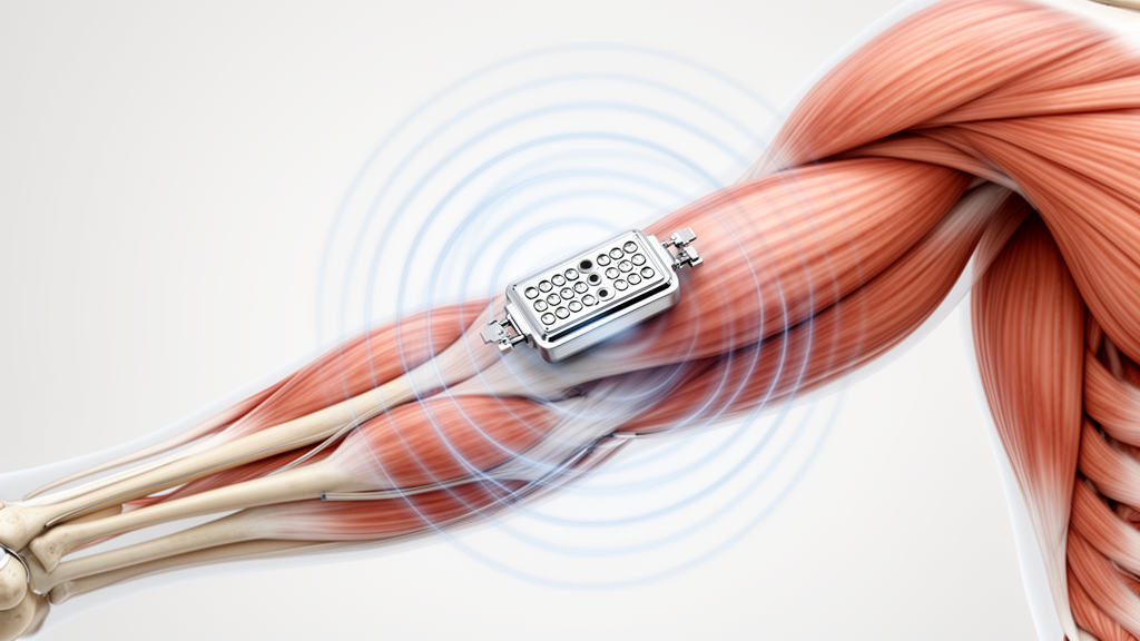
A perspective review published in Frontiers in Physiology examines how magnetomyography, a contactless biomagnetic sensing technique, could provide electrophysiological insights into skeletal muscle integrity to assist future research into sarcopenia. The authors, led by Haijun Li, Zhihao Yao, Yuankun Qi, and Hongyu Zhang from institutions including Shandong University and Beihang University, emphasize that while magnetomyography has made significant technological strides, direct empirical evidence in individuals diagnosed with sarcopenia is currently absent. Sarcopenia is defined clinically as a progressive and generalized skeletal muscle disorder marked by the loss of muscle strength, reductions in muscle quantity or quality, and impaired physical performance. The condition is associated with severe adverse health outcomes, including physical disability, increased fall frequency, bone fractures, and elevated mortality. Despite the growing global health burden of sarcopenia and an estimated annual hospitalization expense of approximately $2,316 per affected patient in the United States, current clinical practice relies heavily on functional assessments that provide limited visibility into the underlying neuromuscular alterations that often precede overt physical decline.
Contemporary diagnostic frameworks, such as the revised European consensus known as EWGSOP2 and the Asian Working Group for Sarcopenia 2019 guidelines, place muscle weakness at the center of clinical evaluation. Under the EWGSOP2 framework, documented low muscle strength identifies probable sarcopenia, confirmed reductions in muscle quantity or structural quality establish a formal diagnosis, and poor physical performance indicates severe disease manifestation. Standard clinical tools including handgrip dynamometry, the chair stand test, gait speed measurement, and the Short Physical Performance Battery are well-validated and practical. However, these tests capture integrated behavioral performance rather than isolated neuromuscular electrophysiology, leaving test scores vulnerable to confounding factors such as patient motivation, localized joint pain, cognitive status, balance disturbances, and participant compliance. Although imaging modalities like computed tomography and magnetic resonance imaging remain gold standards for accurately differentiating fat infiltration from soft tissue, changes in muscle mass alone fail to explain functional decline, particularly in individuals older than 75 years where muscle strength declines far more rapidly than total muscle volume.
To bridge the methodological gap between clinical functional testing and direct neuromuscular evaluation, researchers have investigated electrophysiological tools. Conventional surface electromyography measures electrical potentials at the skin surface, but its signals are substantially distorted by skin-electrode contact impedance, tissue conductivity variations, and volume conduction effects. High-density surface electromyography and invasive needle electromyography offer greater precision, yet they are not routinely integrated into standard sarcopenia diagnostic workflows. Magnetomyography, abbreviated as MMG in this context, offers an alternative modality by non-invasively capturing the ultra-weak biomagnetic fields produced by the ionic currents of depolarizing muscle fibers. The review authors explicitly clarify that this biomagnetic technique must not be confused with mechanomyography, which historically shared the identical abbreviation while tracking mechanical surface vibrations during muscular contractions. Because the magnetic permeability of biological tissue is effectively identical to that of free space, biomagnetic fields propagate through subcutaneous tissue, fat, and skin with minimal conductive distortion, creating potential opportunities for improved spatial localization.
Biomagnetic detection operates according to the classical Biot-Savart law, which dictates that electrical currents coursing through active biological tissues inherently generate surrounding magnetic vector fields. In contracting skeletal muscle, action potentials propagating along individual muscle fibers generate both intracellular and extracellular current elements that produce magnetic fields exhibiting normal and tangential components relative to the skin surface. These generated signals, however, are exceptionally faint, typically registering in the femtotesla to picotesla range, which is several orders of magnitude smaller than the background geomagnetic field of Earth. Historically, recording such minute signals required superconducting quantum interference devices, or SQUIDs, first demonstrated for human skeletal muscle by Cohen and Givler in 1972 using single-channel recordings during isometric contractions of the upper arm and thenar muscles. While modern multichannel SQUID systems deliver femtotesla-level sensitivity, their mandatory reliance on complex cryogenic cooling systems severely limits their portability, operational flexibility, and practical feasibility in routine clinical environments.
Recent technological developments have increasingly shifted toward optically pumped magnetometers, specifically devices operating in or near the spin-exchange relaxation-free regime. Spin-exchange relaxation-free magnetometers utilize the zero-field magnetic resonance of spin-polarized rubidium atoms, achieving theoretical sensitivities that rival or exceed traditional cryogenic SQUIDs while operating entirely at room temperature. The compact footprint of these atomic sensors permits flexible, body-conforming sensor positioning, enabling researchers to configure wearable arrays that measure electrophysiological activity with millisecond temporal resolution while participants move naturally. Furthermore, multi-axis optically pumped magnetometers can measure up to three orthogonal spatial components of the magnetic field simultaneously. This vector capability allows investigators to record directional field distributions that help model source orientation and trace action-potential propagation paths across muscle tissue more accurately than scalar surface voltage maps.
Experimental precedents have demonstrated the technical viability of biomagnetic recording across diverse anatomical structures. Early proof-of-concept investigations utilizing superconducting quantum interference device arrays for reproductive assessment recorded spatiotemporal magnetic signals from uterine contractions and pelvic-floor musculature. Subsequent investigations deploying optically pumped magnetometers confirmed the ability to record evoked compound muscle action potentials from small intrinsic hand muscles following median and ulnar nerve stimulation, as demonstrated in a single-subject evaluation by Broser and colleagues. Later biophysical modeling and multi-sensor recordings identified distinct phases in the magnetomyography waveform, showing that early signal components correspond directly to propagating muscle action potentials, whereas later phases appear linked to activation of the contractile apparatus. Furthermore, work by Noury and colleagues in 2025 demonstrated that independent component analysis could decompose single motor-unit firing patterns from magnetomyography recordings, a capability verified through concurrent intramuscular electromyography that offers a non-invasive window into motor-unit remodeling.
Beyond motor-unit decomposition, magnetomyography has demonstrated sensitivity to fatigue-related neuromuscular adaptations, which represent an important dimension of functional deterioration in sarcopenia. In an experimental protocol evaluating sustained isometric contractions of the rectus femoris in healthy adults, Sometti and colleagues confirmed that optically pumped magnetometers successfully detected fatigue-induced spectral shifts, demonstrating strong concordance with conventional surface electromyographic fatigue metrics. In a longitudinal evaluation involving healthy participants over four weeks of resistance training, Brümmer and colleagues documented the sensitivity of triaxial optically pumped magnetometers to exercise-induced neuromuscular adaptations. Additionally, proof-of-concept testing in three individuals diagnosed with neuromuscular disorders conducted by Semeia and colleagues demonstrated reduced spectral power and altered signal variability relative to healthy control subjects, confirming that biomagnetic sensors can register pathological alterations in neuromuscular organization, despite constraints imposed by small sample sizes and limited sensor bandwidth.
Synthesizing these technological capabilities, the review authors propose a prospective, conceptual assessment framework tailored to investigate sarcopenia mechanisms across clinical research settings. The conceptual model suggests evaluating upper-limb musculature, such as hand intrinsic muscles or forearm flexors to complement handgrip dynamometry, alongside lower-limb targets like the rectus femoris, tibialis anterior, or gastrocnemius to contextualize mobility and gait speed. The recommended assessment protocol incorporates maximal voluntary contractions to determine peak amplitude and motor-unit recruitment capacity, sustained submaximal isometric contractions at thirty to fifty percent of maximal voluntary force to observe spectral median frequency shifts during fatigue, and baseline resting recordings to evaluate spontaneous electrical activity or fasciculation patterns. Derived metrics, including root-mean-square amplitude, power spectral distributions, decomposed motor-unit firing rates, and vector spatial field maps, would be evaluated against established functional markers, bioelectrical impedance analysis, and ultrasound imaging to track interventions.
Despite the conceptual promise of biomagnetic muscle evaluation, the authors outline major physical, methodological, and infrastructural hurdles that must be resolved prior to clinical translation. High-sensitivity atomic magnetometers operating in the spin-exchange relaxation-free regime require heavily attenuated background magnetic fields, historically mandating expensive, stationary magnetically shielded rooms that pose substantial space and cost barriers for healthcare facilities. While portable, cylinder-shaped magnetic shields developed by Iwata and colleagues and mobile mu-metal shielding systems evaluated by Nordenström and colleagues demonstrate that filtering and signal averaging can yield qualitative muscle waveforms outside shielded rooms, these portable setups remain small-scale proofs of concept tested in only a few healthy participants. Furthermore, multi-sensor optically pumped magnetometer arrays remain costly, and operational considerations such as long-term calibration stability, sensor drift, and maintenance requirements continue to limit practical deployment in geriatric care settings.
A critical challenge highlighted in the review involves physiological signal interpretation in older, sarcopenic populations. The net magnetic field recorded at the sensor surface represents a complex superposition of propagating action potentials, motor-unit discharge rates, contractile geometry shifts, and motion artifacts. In sarcopenia, where progressive muscle atrophy, fibrous tissue replacement, denervation, and intramuscular fat accumulation occur concurrently, variations in sensor-to-source distance and tissue geometry can alter measured root-mean-square amplitude independently of neural drive. As demonstrated methodologically by Yang and colleagues, physical changes in source-to-sensor spacing constitute major measurement confounders that require rigorous compensation. The authors conclude that magnetomyography currently lacks normative reference databases across age and sex cohorts, validated test-retest reliability data in frail populations, and direct clinical trials in sarcopenic patients, necessitating interdisciplinary collaboration across geriatrics, physiology, and engineering to establish whether biomagnetic sensing can evolve into a validated clinical diagnostic tool.
Subject of Research: Feasibility and physiological relevance of magnetomyography for skeletal muscle assessment in sarcopenia
Article Title: Magnetomyography for skeletal muscle function assessment: prospective applications in sarcopenia
Article References: Li, H., Yao, Z., Qi, Y., & Zhang, H. (2026). Magnetomyography for skeletal muscle function assessment: prospective applications in sarcopenia. Frontiers in Physiology, 17, Article 1889632. https://doi.org/10.3389/fphys.2026.1889632
Image Credits: AI Generated
DOI: 10.3389/fphys.2026.1889632
Keywords: magnetomyography, sarcopenia, optically pumped magnetometers, skeletal muscle, neuromuscular aging, electrophysiology, biomagnetism, muscle function assessment, skeletal, muscle, function, assessment
News Source: Drew Townsend. (November 12, 2026). Magnetomyography Shows Potential for Evaluating Skeletal Muscle Changes in Sarcopenia Research. Scienmag.