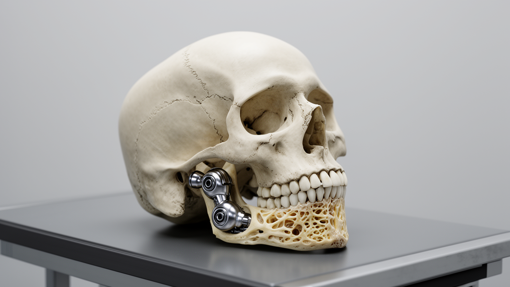
Surgeons have demonstrated that a carefully staged protocol using autogenous bone grafts can successfully support a standard commercial temporomandibular joint prosthesis in a severe case of hemifacial microsomia lacking a natural glenoid fossa. Hemifacial microsomia represents the second most common congenital facial disorder following cleft lip and palate, though its precise etiology remains unknown. In its most severe anatomical presentations, classified under Kaban’s modification of the Pruzansky system as type IIB or type III, patients experience condylar dysplasia or the complete absence of the condyle, glenoid fossa, and mandibular ramus. Restoring facial symmetry and masticatory function in these severe categories typically requires customized, patient-specific total joint replacement systems. However, because customized alloplastic prostheses are subject to prohibitive manufacturing expenses and remain unapproved by regulatory bodies such as national food and drug administrations in several regions, clinicians face substantial technical obstacles when attempting to deploy off-the-shelf commercial hardware.
The central mechanical dilemma in treating severe hemifacial microsomia with stock total joint replacement systems lies in the extensive lack of supporting skeletal framework on the affected cranium and jaw. In individuals classified with type IIB defects, the posterior zygomatic arch and glenoid fossa are completely absent, while the ascending mandibular ramus is exceptionally thin, severely reduced in vertical height, and sharply inclined toward the midline. Stock total joint replacement designs, such as the Biomet system manufactured by Zimmer Biomet Holdings, require solid, stable cortical bone surfaces for anatomical positioning and rigid screw fixation of both the superior temporal fossa component and the inferior mandibular ramal component. Without adequate bone stock and proper angular orientation, securing the fossa plate to an abnormally canted temporal slope is impossible, and seating a preformed mandibular prosthesis fails to achieve symmetrical facial projection, leaving patients with persistent malocclusion and striking mandibular asymmetry.
To overcome these biomechanical barriers, oral and maxillofacial reconstructive surgeons at Seoul National University Dental Hospital devised a multi-phase reconstructive pathway for an 18-year-old male diagnosed with left-sided hemifacial microsomia type IIB. The clinical investigation, formally approved by the hospital’s Institutional Review Board under protocol number ERI 25024, addressed an intricate constellation of structural anomalies on the patient’s left side. Initial physical and radiological evaluations revealed the complete absence of the left temporomandibular condyle, posterior zygomatic arch, and glenoid fossa, alongside an atrophied, foreshortened mandibular body, a thin ascending ramus displaying severe medial inclination, pronounced maxillary canting, and severe mandibular retrognathism accompanied by facial asymmetry. The patient had previously undergone surgical interventions at an external hospital involving an autogenous rib cartilage graft for ear reconstruction and an iliac bone graft designed to augment the left mandibular body, but clinical imaging confirmed that the grafted iliac bone had almost entirely resorbed over time.
The initial phase of the staged surgical management prioritized correcting the severe occlusal plane distortion and maxillary canting through distraction osteogenesis. The surgical team executed a Le Fort I osteotomy across the maxilla combined with a unilateral sagittal split ramus osteotomy on the left side of the mandible. To establish a controlled rotation center during distraction, a thin two-hole miniplate was affixed to the contralateral right zygomaticomaxillary buttress, while an external distraction device was anchored directly to the left maxillary segment. Following temporary intermaxillary fixation to evaluate and test the planned downward vector of the maxilla and the mobilized distal mandibular segment, the distraction appliance was activated after a one-week latency period. Bone elongation proceeded at a rate of one millimeter per day over seven consecutive days, achieving an overall vertical correction of seven millimeters and successfully leveling the maxillary cant.
During this first distraction phase, the reconstructive team observed an important mechanical limitation inherent to the anatomical deformity of hemifacial microsomia. Because the left proximal mandibular segment entirely lacked native temporomandibular joint components, including a fibrous capsule and stabilizing articular ligaments, it could not provide sufficient physiological resistance against the simultaneous downward vector exerted on the distal jaw segment during distraction. Consequently, the proximal bone moved downward in tandem with the distracted distal segment, an effect confirmed postoperatively by the complete absence of a conventional bony step at the antegonial notch. Although distraction successfully elongated both the maxilla and mandible while eliminating the occlusal cant, the underlying deficiency in bone thickness, the medial ramal tilt, and the absent glenoid fossa still required targeted structural remediation before any prosthetic hardware could be implanted.
Six months following the completion of distraction osteogenesis, the surgeons carried out a second major operative procedure centered entirely on autogenous bone grafting to reconstruct a functional anatomical platform. To resolve the transverse ramal discrepancy and thin cortical dimensions, a unilateral sagittal split ramus osteotomy was repeated on the left mandible. The clinicians harvested an autogenous block graft from the patient’s iliac crest and interposed it between the separated proximal and distal osteotomy segments. This intersegmental wedge graft laterally transposed the proximal bone plate, increasing the overall thickness of the ascending ramus and reducing its abnormal medial tilt, which yielded a measured improvement of approximately 6.5 degrees in the frontal ramal angle. This angular correction established the necessary axial alignment and physical width required to accommodate the mandibular ramal prosthesis in full frontal symmetry with the unaffected right side.
Simultaneously during this second operation, the surgical team engineered a horizontal bony shelf upon the left temporal bone to enable the stable anchorage of the temporal fossa component of the stock joint. Because the natural glenoid fossa was missing, the native temporal surface presented a steep, irregular osseous slope unsuitable for flat plate fixation. To construct a viable recipient site, the surgeons harvested autogenous cortical bone blocks from the patient’s contralateral right mandibular ramus. Three pieces of harvested ramal bone were shaped, layered in an overlapping pyramidal configuration, and secured to the temporal slope using a central mini-screw to create a flat horizontal platform. The residual structural void between the layered bone construct and the sloping temporal bone was filled with an alloplastic bone substitute matrix soaked in 0.5 cubic centimeters of recombinant human bone morphogenetic protein 2, designated rhBMP-2, to facilitate complete osteoinduction and osseous consolidation.
Eight months after the grafting intervention, comprehensive radiological evaluation demonstrated that the transplanted autogenous bone grafts in both the left mandibular ramus and the left temporal bone had achieved complete osseous fusion and stable incorporation. The reconstructive team then proceeded with the primary corrective operation, executing comprehensive two-jaw orthognathic surgery combined with the immediate placement of the stock Biomet total joint replacement system. This third surgical stage incorporated a Le Fort I osteotomy, a unilateral sagittal split ramus osteotomy on the contralateral right mandible, and a simultaneous osseous genioplasty to address residual retrognathism and asymmetric chin deviation. Following intraoperative intermaxillary fixation with a fabricated surgical wafer to lock the dental arches into target occlusion, the two components of the stock alloplastic joint were precisely adapted to the reconstructed skeletal beds.
The installation of the stock Biomet prosthesis required meticulous surface recontouring to optimize direct bone-to-implant contact along the previously grafted bone platforms. The prosthetic fossa component was initially anchored to the reconstructed temporal platform using two fixation screws, after which the metallic condylar ramal component was manually guided into congruence with the prosthetic fossa pocket and secured to the lateral mandibular ramus with two preliminary screws. The surgical team then released the intermaxillary fixation to evaluate dynamic dental occlusion and verify the passive centration of the artificial condyle within the fossa cup. Once optimal articular seating and occlusal relations were established, rigid fixation was finalized by driving two additional screws into the temporal fossa plate, totaling four screws, and three supplementary screws into the mandibular component, totaling five screws across the ascending ramus.
Postoperative lateral cephalometric measurements documented major structural improvements resulting from the combined orthognathic realignment and joint implantation. The analysis recorded a 0.13-degree increase in the sella-nasion-A point angle, designated SNA, alongside a 4.03-degree reduction in the sella-nasion-B point angle, designated SNB, and a 6.59-degree reduction in the overall mandibular plane angle. Crucially, the intervention achieved an anterior advancement of the pogonion measuring 12.42 millimeters, substantially restoring chin projection and harmonizing the profile. Five months following the total joint replacement surgery, the clinicians performed a final minor contouring operation to eliminate residual soft-tissue and skeletal asymmetry, which involved removing provisional osteosynthesis plates, refining the genioplasty, performing a right mandibular angle reduction ostectomy, and placing targeted bone grafts to smooth residual contours.
The patient achieved stable dental occlusion and restored jaw function throughout subsequent postoperative orthodontic therapy, with functional temporomandibular joint dynamics and facial symmetry successfully preserved over a seven-year clinical follow-up period. While extended custom-manufactured temporomandibular joint prostheses are frequently advocated in contemporary craniofacial literature for complex skull base and mandibular defects, their clinical utility remains constrained globally by steep manufacturing costs and national regulatory barriers. The authors emphasize that because this report details the treatment of a single clinical subject, the findings cannot be generalized to all patients presenting with hemifacial microsomia without a glenoid fossa, and larger multi-center cohorts are necessary to fully validate the broader reproducibility of the protocol. Nonetheless, this clinical case confirms that combining staged distraction osteogenesis, intersegmental iliac grafting, and ramal bone grafting with osteoinductive rhBMP-2 provides a reliable surgical strategy for anchoring standard stock total joint prostheses in severely deficient anatomical environments.
Subject of Research: Surgical reconstruction and stock total joint replacement in adult hemifacial microsomia
Article Title: Total joint replacement with the stock Biomet system in adult hemifacial microsomia without glenoid fossa: a case report and literature review
Article References: Yu, K. H., Han, J. J., & Hwang, S. J. (2025). Total joint replacement with the stock Biomet system in adult hemifacial microsomia without glenoid fossa: a case report and literature review. Maxillofacial Plastic and Reconstructive Surgery, 47(1), Article 32. https://doi.org/10.1186/s40902-025-00478-5
Image Credits: AI Generated
DOI: 10.1186/s40902-025-00478-5
Keywords: hemifacial microsomia, total joint replacement, Biomet system, glenoid fossa reconstruction, temporomandibular joint, distraction osteogenesis, sagittal split ramus osteotomy, autogenous bone graft, iliac bone graft, rhBMP-2, orthognathic surgery, mandibular asymmetry, frontal ramal angle, Le Fort I osteotomy, pogonion advancement, maxillofacial surgery
News Source: Ophelia Keating. (November 20, 2026). Staged Bone Grafts Enable Stock Joint Implants in Hemifacial Microsomia. Scienmag.