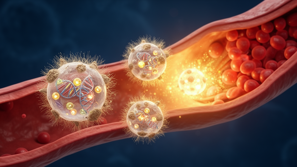
Doxorubicin, a widely used anticancer drug, triggers a dose-dependent cardiotoxicity that can progress into irreversible, progressive myocardial injury and heart failure, and there is currently no standard therapeutic strategy for this condition. Now, a research team from Dalian Medical University has engineered a strikingly clever delivery vehicle: a nanoparticle wrapped in the membrane of a macrophage, an immune cell that naturally homes in on inflamed tissue. Inside this biomimetic shell, the researchers packed a two-pronged payload — celastrol, a natural anti-inflammatory compound, and small interfering RNA that silences a gene driving cardiac cell death. In mice with doxorubicin-induced cardiomyopathy, the system restored cardiac function, curbed fibrosis, and protected mitochondria, offering a detailed preclinical demonstration of how immune-cell camouflage could carry combination therapy directly to a damaged heart.
The clinical problem the team set out to solve is genuinely unmet. Doxorubicin-induced cardiomyopathy, or DIC, begins with inflammatory cell infiltration into the myocardium and evolves through myocardial fibrosis, oxidative stress, mitochondrial dysfunction, and DNA damage, ultimately killing cardiomyocytes. Dexrazoxane is the only approved therapeutic option, and it works by chelating intracellular ferrous iron to block Fenton-reaction-mediated ferroptosis — an iron-dependent form of programmed cell death driven by lipid peroxidation. Yet its true efficacy remains controversial because of significant side effects. Doxorubicin disrupts iron homeostasis by modulating iron-regulatory proteins, depleting the ferroptosis defenders GPX4 and glutathione, while iron accumulation simultaneously activates pro-inflammatory signaling. This entanglement of inflammation and ferroptosis is precisely why the researchers pursued a dual-mode strategy attacking both processes at once.
The inflammatory arm of the attack centers on ADAM17, a transmembrane protease that sheds the extracellular domains of numerous membrane-bound factors and activates multiple downstream pathways. Recent work confirmed that ADAM17 exacerbates DIC by promoting TNF-α release, upregulating TRAF3, enhancing TAK1 phosphorylation, and finally activating the MAPK signaling cascade that induces cardiomyocyte apoptosis. In mice with cardiac-specific ADAM17 knockout, cardiac dysfunction, fibrosis, and apoptosis are all significantly reduced. Silencing ADAM17 with siRNA is highly specific in principle, but siRNA in the bloodstream faces brutal bottlenecks: low delivery efficiency, poor bioavailability, rapid degradation by nucleases, and a short circulation time. Celastrol, the complementary arm, is a pentacyclic triterpenoid with anti-inflammatory, antioxidant, and cardioprotective credentials in atherosclerosis, valve disease, and heart failure — but its poor water solubility and low bioavailability have stalled clinical translation, and its effects in chronic DIC were rarely reported.
The delivery platform that unites these payloads is a feat of modular nanoengineering. The team first synthesized HA-TK-PEG-Chol, a reactive-oxygen-species-responsive amphiphilic molecule built on hyaluronic acid, a non-toxic, biocompatible, non-immunogenic glycosaminoglycan. This carrier self-assembles in PBS and encapsulates celastrol via nanoprecipitation, forming the HC nanocarrier. In parallel, a positively charged lipid complex of DOTAP and DOPE binds negatively charged siADAM17 through electrostatic interactions, forming the DA carrier; agarose gel electrophoresis identified an optimal nitrogen-to-phosphorus ratio of 4:1. Mixing HC with DA yields HCDA, and coating that particle with an equal mass of macrophage membrane — isolated from RAW 264.7 cells, sonicated, and co-extruded through 1000 nm and then 200 nm polycarbonate membranes — produces the final biomimetic nanoparticle, M@HCDA.
Characterization confirmed the construction in vivid detail. Western blotting showed the membrane fraction expressed the macrophage markers F4/80 and CD68 while lacking cytoplasmic and nuclear contaminants. Dynamic light scattering measured M@HCDA at 157.67 ± 4.04 nm with a polydispersity index of 0.12, and its zeta potential settled at 7.4 ± 0.38 mV. Transmission electron microscopy revealed a distinct light-colored membrane-like coating over dark, dense interior particles, and Coomassie Brilliant Blue staining showed M@HCDA protein band patterns matching the macrophage membrane. Encapsulation efficiencies reached 77.0 percent for celastrol and 72.3 percent for siADAM17 in the coated formulation. Most importantly, the membrane cloak dramatically improved stability: over seven days of storage and in 10 percent serum, particle size and polydispersity barely changed, while uncoated HCDA fluctuated and aggregated. Naked siADAM17 degraded almost immediately upon exposure to serum, forming siRNA-protein complexes within 10 minutes, whereas M@HCDA delayed complex formation to 20 minutes — evidence that the membrane forms a natural barrier against nuclease invasion and serum protein adsorption.
In vitro, the payload proved both safe and potent. Across mouse HL-1 atrial muscle cells and human AC16 cardiomyocytes, M@HCDA caused no significant cytotoxicity at any tested dose, with Live/Dead staining confirming negligible cell death. When cells were injured with 1 μM doxorubicin — the concentration that cut viability to roughly 50 percent — M@HCDA improved survival in a dose-dependent manner, outperforming single-mode formulations and even the uncoated HCDA composite. Mechanistic staining told the story: doxorubicin sharply elevated reactive oxygen species and ferrous iron, and M@HCDA significantly suppressed both. C11-BODIPY staining revealed massive lipid peroxidation in injured cells, which the treatment reversed — and the HC component outperformed the DA component in inhibiting iron accumulation and peroxidation markers, indicating the anti-ferroptosis effect was primarily attributable to celastrol.
Targeting experiments showed the macrophage cloak earning its keep. Fluorescent nanoparticles loaded with coumarin 6 were taken up by injured HL-1 cells in a time-dependent fashion, with the membrane-coated version consistently outperforming the uncoated control at every time point — a result replicated in AC16 cells and neonatal rat cardiac fibroblasts. In living mice traced with the near-infrared dye DIR, ex vivo cardiac imaging at 6, 12, and 24 hours after injection showed the targeted formulation accumulating far more fluorescence in injured hearts than uncoated particles or free dye. Safety profiling was equally reassuring: hemolysis stayed below 5 percent at concentrations under 200 μg/mL, and tail-vein doses up to 400 μg/kg produced normal electrocardiograms, intact organ histology, and unchanged liver and kidney markers including AST, ALT, BUN, and creatinine.
The decisive test came in a chronic mouse model, where doxorubicin was injected intraperitoneally at 5 mg/kg once weekly for four weeks, followed 24 hours later by tail-vein treatments. Doxorubicin alone drove significant ST-segment elevation, impaired diastolic function, and steep declines in ejection fraction and fractional shortening. Both low and high doses of M@HCDA reversed these abnormalities more completely than any comparison group, while slashing serum injury markers CK-MB, cTnI, and LDH toward baseline. Histology matched function: Masson, Sirius Red, and α-SMA staining showed the strongest suppression of interstitial and perivascular collagen deposition, wheat germ agglutinin staining revealed cardiomyocyte cross-sectional areas restored to near-healthy size, and TUNEL co-staining with the cardiomyocyte marker TNNT2 found very few apoptotic cells in treated hearts. Strikingly, the component controls proved the synergy: macrophage membrane, free celastrol, and empty carrier each failed to improve cardiac function, and the celastrol group’s survival fell to just 20 percent, likely because the insoluble drug injected in PBS exacerbated cardiac load.
Mechanistic dissection tied everything together. RNA sequencing of myocardial tissue identified 206 co-expressed genes through which M@HCDA appears to act, with Gene Ontology and KEGG analyses showing that doxorubicin upregulated collagen fibril organization, iron ion homeostasis, inflammatory cell apoptosis, mitochondrial depolarization, and the ferroptosis and MAPK signaling pathways — all of which were downregulated after treatment, while the TCA cycle, fatty acid metabolism, and AMPK pathway rebounded. Molecular docking predicted celastrol binding to GPX4 at −7.312 kcal/mol and to SLC7A11 at −8.904 kcal/mol, and molecular dynamics simulations showed the complexes more stable than the protein monomers, with a single deep energy well indicating sustained low-energy conformations. Western blots confirmed that treated hearts recovered GPX4 and SLC7A11 while suppressing phosphorylated TAK1 and P38, and Mito-Tracker and JC-1 staining plus bio-electron microscopy showed mitochondrial membrane potential restored and cristae preserved. Four weekly doses of M@HCDA in healthy mice caused no chronic toxicity. Together, these preclinical results demonstrate that a macrophage-mimicking nanoparticle can co-deliver a gene drug and a natural cardioprotective compound to the injured myocardium in mice, silencing ADAM17 to calm inflammation while celastrol stabilizes the ferroptosis machinery — a multi-target synergy that opens new experimental avenues for protecting the heart against chemotherapy injury, pending the translational work that any mouse study requires.
Subject of Research: Targeted co-delivery of celastrol and siADAM17 via macrophage biomimetic nanoparticles for doxorubicin-induced cardiomyopathy therapy in mice
Article Title: Targeted delivery of celastrol and siRNA via macrophage biomimetic nanoparticles for doxorubicin-induced cardiomyopathy therapy
Article References: Zhang, Q., Guo, T., Xue, X., Zhang, Z., Liu, X., Cai, Q., Wang, N., Zhang, B., & Peng, J. (2026). Targeted delivery of celastrol and siRNA via macrophage biomimetic nanoparticles for doxorubicin-induced cardiomyopathy therapy. Materials Today Bio, 41, Article 103779. https://doi.org/10.1016/j.mtbio.2026.103779
Image Credits: AI Generated
DOI: 10.1016/j.mtbio.2026.103779
Keywords: doxorubicin-induced cardiomyopathy, biomimetic nanoparticles, macrophage membrane, celastrol, siRNA, ADAM17, ferroptosis, GPX4, TAK1-MAPK pathway, targeted drug delivery, cardiotoxicity, preclinical study
News Source: Nathaniel Bowman. (October 26, 2026). Macrophage-Mimicking Nanoparticles Ferry Gene Drug and Natural Compound to Repair Chemotherapy-Damaged Hearts. Scienmag.