Hearing researchers have long dreamed of cochlear implants that speak to the auditory nerve with the precision of natural sound. A new study in EMBO Molecular Medicine brings that vision closer, showing that multichannel optical cochlear implants, paired with a light-sensitive gene therapy, can drive spectrally distinct activity in the auditory midbrain of gerbils at energy levels that, for the first time, look compatible with clinical devices. The work, led by Niels Albrecht, Elisabeth Koert and Tobias Moser at the University Medical Center Göttingen, demonstrates that individual light-emitting channels on an implant can activate tonotopically ordered, distinguishable populations of auditory neurons more cleanly than the electrical contacts used in today’s implants.
The central problem the team tackled is spectral resolution. Conventional electrical cochlear implants bypass damaged hair cells and stimulate spiral ganglion neurons directly, restoring open speech perception for most users after a year of rehabilitation. But each electrode spreads current broadly through the fluid-filled cochlea, activating large, overlapping populations of neurons.
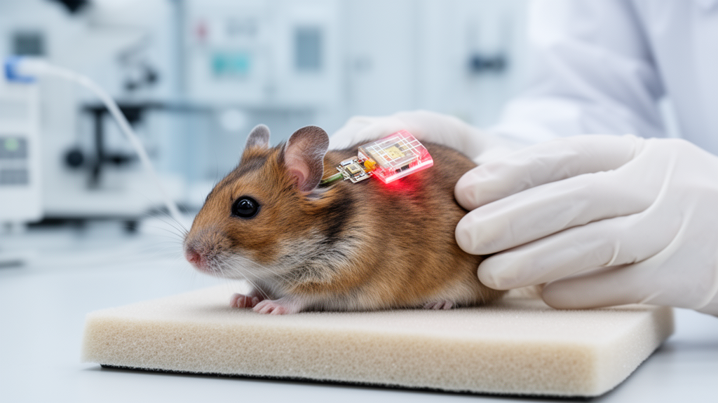
The study, published as an open-access article on 22 October 2026, rests on two technical advances that had previously been pursued separately. The second is a light-based implant built from five or ten individually addressable green LEDs, each measuring 220 by 270 micrometers, mounted on a flexible polyimide substrate at spacings of 300 or 400 micrometers and encapsulated in silicone. By combining the two, the researchers sought to test whether individual optical channels could activate the auditory pathway reliably at radiant energies low enough to be plausible for a battery-powered clinical device.
To make the auditory nerve light-sensitive, the team injected an adeno-associated virus, AAV-PHP.S, carrying the ChReef gene under the control of the human synapsin promoter into the cochleae of Mongolian gerbil pups on postnatal day 8 or 9. After at least eight weeks, they checked whether the gene transfer had worked by placing an optical fiber at the round window and recording optically evoked auditory brainstem responses. Such responses to sub-microjoule light pulses appeared in 13 of 15 treated gerbils, with a median threshold of about 0.32 microjoules. Postmortem immunohistochemistry confirmed that between 24 and 74 percent of spiral ganglion neurons were ChReef-positive in animals with responsive thresholds below 10 microjoules. Notably, the injected cochleae showed a roughly 30 percent lower median spiral ganglion neuron density than untreated controls, a difference that was statistically significant against control animals but not against the untreated contralateral ears of the same animals, and the reduction was strongest in the apical turn. The authors suggest this loss may relate to pressure during cochlear injection and note that alternative viral delivery approaches might preserve neuron density in future work.
The key physiological readout came from the inferior colliculus, a midbrain hub through which nearly all ascending auditory information passes. The researchers inserted a 32-channel linear electrode array along the tonotopic axis of the central nucleus of the inferior colliculus, allowing them to estimate which cochlear frequency regions were being activated by any given stimulus. Complete recordings were obtained from nine of the thirteen gene-treated animals with usable brainstem responses and from eight non-injected control animals, in which acoustic input to the right ear was deliberately blocked to match the stimulated side. Because the treated animals were not deafened, pure tones could be used to map each electrode’s characteristic frequency before implant stimulation began. The measured tonotopic slope of about 4.75 octaves per millimeter matched literature values well.
Once the LED implants were inserted into the cochlea through the round window or a basal-turn cochleostomy, every implanted animal showed reliable inferior colliculus responses to 1-millisecond light pulses from individual emitters, with a median threshold of 0.15 microjoules. This consistency across channels within the same animal addresses a recognized weakness of earlier prototypes, in which the light sensitivity of the channelrhodopsin CatCh frequently forced researchers to drive several LEDs simultaneously, and reliable responses from all channels of a multichannel device were not consistently observed. The new threshold also sits on the same order of magnitude as the roughly 0.05 microjoules used in clinical electrical stimulation, which is central to the authors’ claim that the energy budget is approaching a translationally feasible range.
To compare intensity coding across stimulation types, the team quantified evoked firing rates in a fixed 13-millisecond onset window, a choice made to keep comparisons fair given that the acoustic stimuli were 100-millisecond pure tones while the optical and electrical stimuli were brief pulses. Rate-level functions showed that firing increased with stimulus intensity in all modalities, but single LEDs rarely reached saturation even at the maximum tested output of about 3 milliwatts, meaning the apparent dynamic range of 11.3 decibels likely underestimates what optical stimulation could achieve. Maximum firing rates were highest for acoustic stimulation, with optical and electrical stimulation statistically indistinguishable from each other and both below the acoustic maximum. By mapping the maximal optically evoked firing rate onto the acoustic rate-level function, the researchers estimated that a single LED achieved a median maximal midbrain activity equivalent to pure tones of about 51 decibels sound pressure level, essentially matching the roughly 50 decibels estimated for single electrical electrodes. The authors caution that this remains below conversational speech levels of around 65 decibels and that the comparison is necessarily approximate, since natural sounds distribute energy across frequency bands whereas the estimate reflects a single channel.
The spectral findings form the heart of the paper. Spatial tuning curves constructed from the midbrain recordings showed tonotopically ordered activation by the LED arrays, and the relationship between emitter position and the characteristic frequency of the best-responding electrode yielded a slope of 1.31 millimeters per octave, close to the 1.4 millimeters per octave predicted by the gerbil Greenwood frequency-place function. Postmortem X-ray tomography of explanted cochleae allowed the team to register each LED’s actual position and orientation within the spiral ganglion, and the tonotopic locations of the evoked responses matched the expectations from emitter placement closely.
However, the high light sensitivity of ChReef also exposed a complication: in roughly half of the recordings, stimulation by a single LED produced not just one but two, and occasionally three, spectral peaks of activation. The additional peaks appeared mostly at lower frequencies than the main peak and, according to the tomographic reconstruction, likely arose from light spreading across the cochlear turn to activate neurons on the opposite side of Rosenthal’s canal. The intensity range over which only the main peak was present had a median of 0.60 milliwatts, and the firing rate carried by the main peak alone before any secondary peak appeared reached a median of 272 hertz. The authors interpret these off-target activations as a consequence of using such a sensitive opsin in the small rodent cochlea, and they note that a previous modeling study of the 2.5-fold larger human cochlea did not predict this cross-turn activation, though they emphasize this remains to be tested directly.
Quantifying the spread of excitation across activity levels revealed a clear ordering: acoustic stimulation produced the narrowest activation, electrical stimulation the broadest, and optical stimulation fell in between. At 200 hertz evoked firing rate, the difference between optical and electrical spread reached a maximum of 2.85 octaves, or 57 percent of the electrical value, while optical spread exceeded the acoustic reference by a comparatively stable 0.8 to 1.1 octaves at low-to-moderate levels. When secondary peaks were excluded from the optical analysis, the main peak’s spread tracked the acoustic reference much more closely, demonstrating that off-target activation, not the primary optical channel, drove much of the broadening at higher intensities.
Two complementary analyses then addressed whether distinct channels produce genuinely distinguishable neural activity patterns. Representational similarity analysis, which compares the correlation distance between population response patterns, showed that optical channels organized their responses in a more tone-like manner than electrical electrodes did, with dissimilarity increasing with channel distance and remaining relatively stable across intensity levels, whereas electrical responses were shaped more by intensity than by electrode identity. Linear discriminant analysis, a cross-validated decoding approach, extended this finding: at activity levels of 350 hertz and above, optical channels could be discriminated at significantly smaller inter-channel spacings than electrical channels, with the electrical threshold averaging about 1.7 times the optical threshold across the tested range. The authors note that because electrical thresholds were frequently censored at the upper bound of the sampled spacing range, this difference is likely conservative.
The researchers are explicit about the study’s boundaries. ChReef’s slow closing kinetics, roughly 30 milliseconds at physiological temperature, restrict stimulation rates to about 50 hertz and would need faster opsins for faithful temporal coding. Only single-channel stimulation was tested, leaving channel interactions during simultaneous multichannel activation unexamined. The animals had intact inner hair cells, whereas future patients would not, although prior work suggests deafening does not substantially alter the relevant measures. The electrical comparison data were reanalyzed from a previous study using the same setup and pipeline, but subtle experimenter-related differences cannot be fully excluded. The authors also stress that behavioral studies will be needed to determine whether the improved spectral encoding observed at the physiological level translates into enhanced perceptual discrimination, since the present work measured midbrain activity rather than hearing itself.
Subject of Research: Preclinical testing of multichannel optical cochlear implants with ChReef optogenetic stimulation in gerbils
Article Title: Multichannel optical cochlear implants enable spectrally distinct auditory activity
Article References: Albrecht, N., Koert, E., Vavakou, A., Roos, L., Jablonski, L., Marcoleta, J. P., Cardona Audi, J., Alfken, J., Klein, E., Ruther, P., Mager, T., Kusch, K., & Moser, T. (2026). Multichannel optical cochlear implants enable spectrally distinct auditory activity. EMBO Molecular Medicine. https://doi.org/10.1038/s44321-026-00541-2
Image Credits: AI Generated
DOI: 10.1038/s44321-026-00541-2
Keywords: cochlear implants, optogenetics, ChReef, hearing loss, spiral ganglion neurons, inferior colliculus, spectral resolution, gerbils, gene therapy, LED stimulation, auditory neuroscience, neural prosthetics
News Source: Cassandra Pierce. (October 28, 2026). Light-Based Cochlear Implants Sharpen the Ear’s Frequency Map in Gerbils. Scienmag.