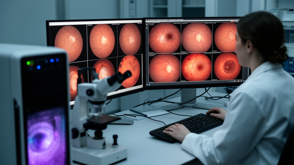
Diabetic retinopathy screening datasets contain too few images of the most serious disease stages for automated classifiers to learn them reliably. A new study shows that a class-conditional latent diffusion model can fill that gap with synthetic retinal fundus photographs, raising validation accuracy across multiple image-classification architectures while preserving the 512 × 512-pixel detail needed to depict small lesions. The researchers developed the Diabetic Retinopathy Latent Diffusion Synthesizer, or DR-LDS, and tested it as an augmentation tool for 11 deep-learning models trained on the APTOS 2019 benchmark. The best reported validation accuracy reached 95.60% with the lightweight SqueezeNet1_1 network, and the study reported statistically significant accuracy gains of up to 15.25% across the evaluated architectures.
The motivation is a familiar problem in medical artificial intelligence, but one with a particularly sharp edge in retinal imaging. APTOS 2019 contains 3,662 fundus images labeled on the five-level International Clinical Diabetic Retinopathy severity scale. Nearly half, 49.3%, were assigned to the “No DR” category, whereas severe and proliferative disease accounted for only 5.3% and 8.1%, respectively. A model trained on such a long-tailed distribution can achieve deceptively high overall accuracy by favoring common cases while performing poorly on the grades that may require urgent intervention. Conventional augmentation with rotations, flips, crops, and brightness or contrast changes increases the number of training examples but does not create new combinations of pathological features.
DR-LDS instead generates images according to a specified disease grade. Its central technical move is to perform diffusion in a compressed latent space rather than directly on pixels. A variational autoencoder first converts each 512 × 512 fundus image into a 64 × 64 × 4 representation, reducing the number of values processed at each diffusion step from 786,432 to 16,384, a 48-fold dimensional reduction. The autoencoder was fine-tuned for 50 epochs on the training portion of APTOS 2019 so that it would retain the fine vessels, microaneurysms, hemorrhages, and exudates that can be smoothed away by a general-purpose compression model. Its weights were then frozen while the diffusion network was trained.
Within that latent space, a conditional U-Net learns to reverse a 1,000-step process that progressively adds Gaussian noise. A separate embedding network converts the diffusion timestep and disease-grade label into a 512-dimensional context vector. That vector is injected into the U-Net through adaptive group normalization and a multi-head cross-attention bottleneck at 16 × 16 spatial resolution. The researchers describe this arrangement as a way to preserve localized, high-frequency pathological detail instead of treating all retinal grades as globally similar images. Skip connections carry fine spatial information through the encoder and decoder, while the attention layer links image features with the requested disease class.
The resulting framework contains 16.36 million parameters and was trained for 150 epochs. The reported training time was 17 minutes and 22 seconds per epoch, although standardized benchmarking was conducted on an NVIDIA A100 GPU. The authors report that the model’s memory footprint is compatible with consumer GPUs having roughly 16 to 24 GB of video memory, but the study did not present a full consumer-device training run. Its estimated weight file occupies about 1.20 GB in full precision, and the denoising process is reported at approximately 34.2 GFLOPs per step. These measurements explain how the method can avoid the computational burden associated with training diffusion models directly in high-dimensional pixel space.
In standardized comparisons against GAN- and diffusion-based baselines retrained on the same APTOS split, DR-LDS achieved a Fréchet Inception Distance of 8.05 and a Kernel Inception Distance of 0.017. Lower values indicate closer resemblance between the distributions of real and generated features. The study reports that the standard DDPM baseline had an FID of 32.38, 256 million parameters, and a training time exceeding one hour per epoch. The authors also report that reducing image resolution from 512 × 512 to 128 × 128 increased FID to 32.45, while sharply reducing the U-Net’s base channel width also degraded generation quality. These ablations indicate that the reported gains depended on both high spatial resolution and sufficient model capacity, rather than on class conditioning alone.
To create the balanced training set, the researchers set a uniform target of 2,000 images for each of the five grades. The model generated 6,338 synthetic images in total, supplementing the existing training examples and bringing every class to that target. Sampling used a guidance scale of 3.0 and 50 denoising-diffusion implicit-model inference steps. The 366-image validation set was isolated before preprocessing or model training and was neither used to fine-tune the autoencoder nor exposed to the diffusion model. Leakage tests compared every synthetic image with every validation image using structural similarity and pixel-wise distance. The maximum SSIM value was 0.42, with a mean of 0.15 ± 0.04, far below the 0.95 threshold used in the study to indicate duplication.
The synthetic images were then used to train 11 convolutional neural-network classifiers under several optimizer and learning-rate configurations. The central result was that adding class-matched synthetic data improved validation accuracy more consistently than adding the same number of classically transformed real images. SqueezeNet1_1 rose to 95.60% validation accuracy and an F1 score of 0.96 in its best configuration. DenseNet121 reached 90.31% in one configuration, compared with a 74.35% baseline, while gains reported for the evaluated architectures ranged from 3.05% to 15.25% and had p-values below 0.001. Classical augmentation produced smaller increases in the six-architecture comparison: for example, SqueezeNet1_1’s F1 score moved from 0.86 to 0.87, compared with 0.96 after generative augmentation. The benefits were not uniform under every hyperparameter setting, and one MobileNet_V2 configuration regressed by 0.84%.
An augmentation-intensity ablation found that doubling minority classes was insufficient, while expanding every class to 5,000 examples slightly reduced accuracy, which the authors interpret as overfitting to the synthetic distribution. Grad-CAM visualizations showed the classifiers attending to regions containing neovascularization, scattered hemorrhages, and exudates, while vessel-map comparisons indicated that synthetic vascular networks preserved branching, tapering, and connectivity. These analyses support the interpretation that the models learned meaningful lesion patterns rather than obvious synthetic artifacts. Still, the work remains a benchmark study: it uses static two-dimensional photographs, does not model disease progression in an individual over time, and has a small hold-out set containing only 28 severe and 22 proliferative cases. The authors consequently describe the results as promising diagnostic potential rather than readiness for unmonitored clinical deployment, and say that larger, multicenter external cohorts would be needed to test performance across populations, cameras, and clinical settings.
Subject of Research: Benchmarking latent diffusion for class-balanced diabetic retinopathy image synthesis and diagnostic-model augmentation.
Article Title: Class balanced diabetic retinopathy image synthesis using a latent diffusion framework
Article References: Alam, T., Liew, T. H., Morol, M. K., Nandi, D., Jubair, M. A.-A., & Rahman, M. (2026). Class balanced diabetic retinopathy image synthesis using a latent diffusion framework. Discover Artificial Intelligence, 6(1), Article 1205. https://doi.org/10.1007/s44163-026-02184-1
Image Credits: AI Generated
DOI: 10.1007/s44163-026-02184-1
Keywords: diabetic retinopathy, latent diffusion, synthetic medical images, class imbalance, fundus photography, deep learning, data augmentation, image synthesis, retinal screening, Class, balanced, diabetic
News Source: Denise Maddox. (October 26, 2026). Latent diffusion balances diabetic retinopathy data and boosts screening models. Scienmag.