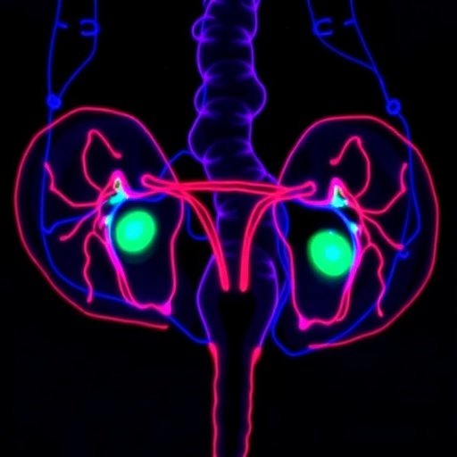
Ovarian cancer may soon be examined through a new kind of microscope that relies on the natural light-emitting properties of biological tissue rather than conventional dyes. A study published in Light: Science & Applications describes a label-free diagnostic approach that combines two-photon autofluorescence microscopy with artificial-intelligence-assisted image processing. The method is designed for histopathological diagnosis, the process by which specialists inspect tissue architecture and cellular features to determine whether cancer is present. Although the available report identifies the imaging strategy and computational framework, it does not provide detailed performance results in the supplied material. Its central premise is nevertheless significant: tissue may be classified by its intrinsic optical signals, potentially reducing the need for staining and making some stages of cancer analysis faster and more reproducible.
Ovarian cancer is particularly difficult to diagnose because its symptoms can be vague, its biological subtypes differ substantially, and malignant tissue may resemble benign or borderline lesions. In routine pathology, tissue removed during surgery or biopsy is typically fixed, embedded, cut into thin sections and treated with chemical stains. These stains reveal nuclei, connective tissue, cytoplasm and other structures, allowing pathologists to interpret the organization of the sample under a conventional microscope. The method remains indispensable, but it is also dependent on preparation quality, staining consistency and expert judgment. A label-free optical system approaches the problem from a different direction. Rather than adding contrast agents, it seeks to measure signals already produced by molecules within the tissue and then uses computational analysis to convert those signals into diagnostically useful maps.
Two-photon microscopy generates those signals by directing ultrashort pulses of near-infrared light into a specimen. In ordinary fluorescence imaging, a molecule absorbs a single photon and then emits light of a longer wavelength. In two-photon excitation, two lower-energy photons arrive at nearly the same time and together provide the energy needed to excite the molecule. Because the probability of this event is extremely low except at the tightly focused point of a laser beam, excitation is naturally confined to a small three-dimensional region. This localization can reduce out-of-focus background and enables optical sectioning, allowing researchers to build depth-resolved images without physically slicing through every layer during observation. Near-infrared light can also penetrate biological material more effectively than shorter wavelengths, although the quality and depth of imaging depend on the tissue and the optical system.
The word “autofluorescence” refers to fluorescence originating from endogenous molecules rather than externally applied dyes. Metabolic cofactors such as NADH and flavin-containing compounds can emit characteristic signals, while structural components including collagen contribute through fluorescence or related nonlinear optical effects. The abundance, chemical environment and spatial distribution of these molecules can change as cells become malignant. Cancer-associated alterations in metabolism, extracellular matrix organization, nuclear structure and cellular density may therefore leave an optical signature. Such signals are not equivalent to a diagnosis on their own; they are measurements that require careful interpretation. The study’s proposed framework addresses that challenge by pairing the microscope with image-processing algorithms intended to improve the clarity of the raw data and identify relevant tissue regions.
The first computational component, described as joint denoising, is aimed at suppressing noise while preserving diagnostically important details. Optical images collected at low signal levels often contain random fluctuations caused by photon statistics, detector electronics, laser instability and background light. Aggressive smoothing can make an image appear cleaner but may erase thin boundaries, small nuclei or subtle texture differences. Denoising algorithms therefore face a balancing problem: they must remove unwanted variation without manufacturing structures that were not present in the specimen. A joint framework implies that denoising is not treated as an isolated cosmetic step. Instead, image restoration is linked to the next task, segmentation, so that the system can preserve features that are useful for separating tissue compartments or identifying cellular patterns.
Segmentation is the process of dividing an image into meaningful regions. In ovarian histopathology, those regions might include nuclei, epithelial structures, stroma, blood vessels, necrotic areas or other compartments that help characterize a lesion. Conventional segmentation can rely on manually chosen thresholds or hand-designed rules, but biological images rarely obey simple boundaries. Cells overlap, tissue textures vary and disease-related changes may be gradual rather than sharply defined. A learned model can be trained to recognize patterns across many examples, producing a pixel-level or region-level map of the image. When denoising and segmentation are optimized together, the system can theoretically use structural information to guide restoration while using cleaner images to improve delineation. That interaction is the technical core of the reported approach.
The potential advantage of combining optical imaging and computation is speed at the interface between measurement and interpretation. A microscope can acquire rich images, but the resulting data may be too complex for a human observer to evaluate efficiently in raw form. An algorithm can quantify intensity, texture, shape and spatial relationships across thousands of image regions, while a segmentation map can focus attention on structures most relevant to diagnosis. In a clinical setting, such tools would not necessarily replace pathologists. More plausibly, they could support review by highlighting suspicious regions, standardizing measurements or helping laboratories compare samples acquired under different conditions. Any such role would require extensive validation against established histopathological diagnoses, testing across institutions and scanners, and careful assessment of errors in both common and rare tumor subtypes.
Label-free imaging also raises practical questions about how a new optical diagnosis would fit into existing workflows. Conventional histology provides a permanent stained record that can be examined repeatedly and archived. Two-photon autofluorescence produces a different kind of information: a map of endogenous optical behavior that may be highly sensitive to preparation, fixation, tissue thickness and imaging settings. Algorithms trained on one instrument or sample protocol may perform less reliably when those conditions change. Standardized acquisition procedures, calibration controls and transparent reporting would therefore be essential. Researchers would also need to determine whether autofluorescence patterns remain stable over time and whether they are specific to ovarian malignancy rather than reflecting inflammation, tissue damage, treatment effects or other noncancerous processes.
The study’s publication signals growing interest in diagnostic systems that unite advanced microscopy with machine learning, but the bibliographic information supplied for this report does not include accuracy values, patient numbers, tumor subtypes, comparison groups or clinical validation outcomes. Those details are crucial for judging whether the technique is ready for practical use. A visually compelling image or an effective laboratory demonstration cannot by itself establish clinical utility. The decisive tests will involve independent samples, blinded evaluation and comparison with expert pathology, alongside measurements of sensitivity, specificity, reproducibility and processing time. If future studies establish that the joint denoising-and-segmentation framework can preserve meaningful tissue features while reducing diagnostic ambiguity, two-photon autofluorescence could become a valuable complement to stained histology. For now, the work presents a technically distinctive route toward label-free ovarian cancer assessment, built on the idea that the tissue’s own light—and algorithms capable of interpreting it—may reveal patterns hidden from conventional inspection.
Subject of Research: Label-free ovarian cancer histopathological diagnosis using two-photon autofluorescence microscopy and joint denoising and segmentation
Subject of Research: Technology and Engineering
Article Title: Label-free ovarian cancer histopathological diagnosis using two-photon autofluorescence microscopy with a joint denoising and segmentation framework
Article References: Pan, Z., Song, N., Cheng, S., Pang, W., Liao, H., Wang, Y., & Gu, B. (2026). Label-free ovarian cancer histopathological diagnosis using two-photon autofluorescence microscopy with a joint denoising and segmentation framework. Light: Science & Applications, 15(1), Article 366. https://doi.org/10.1038/s41377-026-02464-6
Image Credits: AI Generated
DOI: 10.1038/s41377-026-02464-6
Keywords: ovarian cancer, label-free imaging, two-photon microscopy, autofluorescence, histopathology, denoising, image segmentation, artificial intelligence
Cite this news
APA MLA Chicago
SCIENMAG. (August 28, 2026). Label-Free Ovarian Cancer Diagnosis Enhanced by Two-Photon Autofluorescence and Joint Image Processing. https://scienmag.com/label-free-ovarian-cancer-diagnosis-enhanced-by-two-photon-autofluorescence-and-joint-image-processing/
SCIENMAG. “Label-Free Ovarian Cancer Diagnosis Enhanced by Two-Photon Autofluorescence and Joint Image Processing.” Scienmag, 28 August 2026, https://scienmag.com/label-free-ovarian-cancer-diagnosis-enhanced-by-two-photon-autofluorescence-and-joint-image-processing/. Accessed 28 August 2026.
SCIENMAG. “Label-Free Ovarian Cancer Diagnosis Enhanced by Two-Photon Autofluorescence and Joint Image Processing.” Scienmag. August 28, 2026. https://scienmag.com/label-free-ovarian-cancer-diagnosis-enhanced-by-two-photon-autofluorescence-and-joint-image-processing/
Copy citation Download RIS
Tags: advances in microscopic imaging for oncologyAI-assisted image processingAI-assisted image processing in pathologycomputational analysis of tissue imagescomputational frameworks in medical imagingdigital pathology and image classificationhistopathological cancer detectionhistopathological tissue analysisimprovements in histopathology methodsintrinsic optical signals in tissueintrinsic optical signals in tissueslabel-free cancer tissue classificationlabel-free ovarian cancer diagnosislabel-free tissue imagingnon-invasive cancer detection techniquesnon-invasive ovarian cancer detectionoptical imaging for cancer diagnosisoptical imaging in cancer diagnosisovarian cancer diagnosisrapid cancer diagnosis techniquesrapid ovarian cancer screening methodstissue architecture analysis without stainstwo-photon autofluorescence microscopy