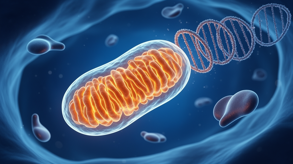
A comprehensive narrative review published in Frontiers in Nutrition examines the complex biological interplay between high-intensity interval training, mitochondrial integrity, and innate immune signaling. The authors synthesize findings across multiple organ systems to propose that the intense metabolic stress generated during high-intensity intervals can destabilize mitochondrial membranes and trigger the leakage of mitochondrial DNA into the cellular cytosol. Once displaced from its normal mitochondrial compartment, double-stranded mitochondrial DNA functions as an endogenous damage-associated molecular pattern that binds to cyclic GMP-AMP synthase, thereby engaging the stimulator of interferon genes pathway. While this signaling axis is well documented in chronic inflammatory and degenerative disease models, the review evaluates whether the transient physiological perturbations of interval exercise engage the same molecular machinery. The authors explicitly characterize their framework as hypothesis-generating, noting that direct in-human confirmation of exercise-induced cytosolic mitochondrial DNA release and distal immune activation remains limited.
High-intensity interval training, characterized by alternating bouts of near-maximal exertion and low-intensity recovery, has gained widespread popularity due to its marked time efficiency and potent capacity to stimulate physiological adaptation. When appropriately dosed, repeated interval bouts enhance mitochondrial oxidative capacity, stimulate tricarboxylic acid cycle flux, improve whole-body glucose homeostasis, and activate key metabolic energy sensors like adenosine monophosphate-activated protein kinase. These positive adaptations are mediated in part by a controlled, dose-dependent rise in reactive oxygen species that stimulates endogenous antioxidant defenses, including superoxide dismutase. However, the authors emphasize that interval protocols frequently lack standardized operational definitions of intensity and dose matching across studies. When training volume or intensity exceeds an individual’s physiological and energetic reserves, excessive reactive oxygen species production shifts the intracellular balance toward oxidative damage. This excessive metabolic strain compromises organelle integrity and establishes the initial cellular conditions under which mitochondrial DNA can escape into the surrounding cytoplasm.
To explain how cellular stress translates into either beneficial adaptation or tissue damage, the review outlines a kinetic, double-threshold model governing cyclic GMP-AMP synthase and stimulator of interferon genes activation. Under this model, an initial adaptive threshold is crossed when moderate mitochondrial DNA release recruits the cytosolic sensor and initiates trafficking of the stimulator protein to the endoplasmic reticulum-Golgi intermediate compartment. In this physiological range, the pathway initiates an autophagic program independent of TANK-binding kinase 1 and interferon production that actively clears misplaced cytosolic DNA, alongside a brief, self-limiting interferon regulatory factor 3 response that supports mitochondrial quality control and immunosurveillance. Conversely, a second, maladaptive threshold is reached when severe metabolic stress causes mitochondrial DNA leakage to overwhelm cellular clearance capacity. Downstream outputs occur with distinct kinetics: early interferon induction begins at the Golgi apparatus, whereas nuclear factor-kappa B activation occurs only with delayed kinetics after the stimulator exits the Golgi for late endolysosomes, transforming unresolved signaling into chronic, non-resolving inflammation.
To maintain rigorous scientific standards, the authors implement an evidentiary grading system across their analysis, classifying source data into three transparent tiers. Tier A comprises direct exercise measurements collected during human or animal training protocols, such as exercise-induced circulating cell-free nucleic acids. Tier B encompasses established mechanistic evidence derived from non-exercise disease, cellular, or toxicological models, while Tier C represents hypothesis-driven extrapolation to exercise physiology. Crucially, the review distinguishes between the intracellular cyclic GMP-AMP synthase pathway and endolysosomal Toll-like receptor 9 signaling. While the cytosolic synthase detects misplaced double-stranded DNA leaking directly from compromised internal mitochondria, Toll-like receptor 9 recognizes unmethylated cytosine-phosphate-guanine motifs within extracellular mitochondrial DNA that has entered the bloodstream and undergone cellular endocytosis. The authors note that other sensors, including absent-in-melanoma 2 and mitochondrial antiviral-signaling proteins, may also converge on shared inflammatory pathways, underscoring that attributing exercise adaptations strictly to the synthase requires cautious interpretation.
In evaluating the central nervous system, the authors emphasize that direct empirical evidence demonstrating interval-induced cytosolic mitochondrial DNA release or pathway activation within human brain tissue is currently absent. Most existing mechanistic observations originate from neurodegenerative models of Alzheimer’s and Parkinson’s diseases, where sustained organelle dysfunction contributes to progressive pathology. In experimental Alzheimer’s disease paradigms, amyloid-beta accumulation elevates reactive oxygen species and promotes voltage-dependent anion channel 1-mediated mitochondrial DNA leakage into the neuronal cytoplasm. This leakage triggers persistent TANK-binding kinase 1 and interferon regulatory factor 3 phosphorylation, driving the release of inflammatory mediators such as interleukin-6 and C-X-C motif chemokine ligand 10 that provoke reactive microgliosis and astrocyte activation. The authors suggest that strenuous interval training could theoretically generate a sub-threshold neuronal stimulus, or alternatively allow exercise-induced circulating mitochondrial DNA to access perivascular microglia through transient changes in blood-brain barrier permeability, thereby engaging microglial Toll-like receptor 9 rather than neuronal cytosolic sensors.
Within the cardiovascular and respiratory systems, mitochondrial integrity is central to continuous physiological function but remains vulnerable to excessive oxidative stress. Cardioprotective remodeling is a well-established outcome of regular exercise training, yet pathological models of pressure overload and diabetic cardiomyopathy demonstrate that chronic mitochondrial DNA leakage drives cardiomyocyte pyroptosis and shifts cardiac macrophages toward a destructive M1 phenotype, exacerbating fibrosis and ventricular dysfunction. Whether acute interval training elicits comparable local mitochondrial leakage within healthy myocardium remains an open scientific question. In the pulmonary system, mechanical and metabolic strains present a unique physiological setting. During intense exertion, pulmonary capillary transmural pressures can exceed forty millimeters of mercury, transiently stressing the delicate blood-gas barrier. The authors hypothesize that this mechanical deformation could theoretically allow pulmonary mitochondrial fragments to enter the systemic bloodstream or trigger local inflammation, although definitive evidence confirming that the lungs act as a primary source of circulating exercise-derived mitochondrial DNA is currently lacking.
Skeletal muscle represents the primary executor of mechanical work during interval exercise and serves as the most direct site for evaluating metabolic strain. Contraction-induced metabolic demands challenge myocyte mitochondria, generating oxidative stress that can modify nucleic acids. Experimental investigations in aged mice subjected to high-intensity training reveal clear markers of mitonuclear imbalance and activation of the mitochondrial unfolded protein response, although direct cytosolic translocation of mitochondrial DNA was not quantified in those models. In exercising humans, strenuous exertion consistently elevates circulating cell-free mitochondrial DNA in plasma, demonstrating that fragments are disseminated into the vascular bed through muscular microcirculation or extracellular vesicles. However, measuring circulating fragments does not establish whether myocyte-specific cytosolic leakage occurred or whether those systemic fragments activate distal immune sensors. Furthermore, endurance training is known to enhance mitochondrial enzymatic activity independently of net alterations in mitochondrial DNA content, illustrating that metabolic adaptations involve nuanced mitochondrial remodeling rather than simple organelle breakdown.
The immune system and metabolic organs, including the liver and white adipose tissue, act as key systemic regulators of interval-induced stress responses. In circulating and tissue-resident macrophages, excessive mitochondrial reactive oxygen species induce abnormal opening of the mitochondrial permeability transition pore, releasing internal nucleic acids that activate the innate synthase cascade and promote pro-inflammatory M1 polarization. In hepatic tissue, lipotoxic challenges, such as excess palmitic acid, stimulate similar organelle pore opening, promoting non-alcoholic steatohepatitis through downstream interferon and nuclear factor-kappa B signaling. The authors highlight documented instances of inter-organ communication, where damaged hepatocytes release extracellular vesicles laden with mitochondrial DNA that travel through permissive sinusoidal barriers to white adipose tissue, triggering local immune sensors and worsening systemic insulin resistance. Although exercise training is recognized for reducing hepatic steatosis and mitigating metabolic inflammation, whether interval regimens directly suppress hepatocyte mitochondrial DNA leakage remains to be formally established through targeted investigation.
Addressing the duality of interval exercise adaptation, the review details a multi-tiered nutritional framework designed to support organelle resilience and modulate innate immune activation across training cycles. Tier 1 strategies focus on upstream mitochondrial protection and metabolic maintenance to prevent organelle destabilization at its initial source. Mitochondrial-targeted antioxidants, including coenzyme Q10 and MitoQ, maintain electron transport chain stability and reduce reactive oxygen species production, supporting membrane potential and potentially lowering the probability of nucleic acid release in high-demand tissues like cardiac and skeletal muscle. Similarly, dietary nitrates, commonly sourced from concentrated beetroot juice, improve mitochondrial oxidative efficiency and contractile economy through nitric oxide signaling pathways. The authors also highlight endogenous beta-hydroxybutyrate, an exercise-inducible ketone body that provides clean cellular fuel, suppresses upstream inflammasome activation, and mitigates mitochondrial-driven inflammatory signaling, although the authors note that ensuring baseline carbohydrate availability represents a primary upstream dietary foundation for preserving energetic homeostasis.
For midstream and downstream modulation, the framework maps targeted phytochemicals and micronutrients while addressing critical translational bottlenecks in sports science. Midstream agents such as phloretin attenuate pore opening via the SHP-2, JAK2, and BAX axis, while isoleucine stabilizes organelle architecture against voltage-dependent leakage. Downstream modulators like rhapontigenin and urolithin A enhance mitophagy clearance through PTEN-induced kinase 1 pathways, while vitamin D and prebiotic-derived short-chain fatty acids foster immunotolerance. However, the authors caution that individual responses to interval training and nutritional interventions are heavily modified by chronological age, baseline training status, sex hormones, and pre-existing insulin resistance. Furthermore, the lack of non-invasive circulating biomarkers to monitor intracellular pathway dynamics poses a significant barrier to clinical implementation. The review strongly advises against indiscriminate high-dose antioxidant supplementation, which can blunt essential hormetic training adaptations, emphasizing that this proposed signaling axis remains a hypothesis-generating model rather than an established clinical prescription.
Subject of Research: Mitochondrial DNA release and cGAS-STING immune signaling during high-intensity interval training
Article Title: Metabolic stress, mitochondrial DNA release and the nutritional regulatory potential of the cGAS-STING pathway during high-intensity interval training
Article References: Xiang, K., Luo, K., Wang, S., Lv, W., Zhou, Z., Li, Y., Wang, R., & Yang, S. (2026). Metabolic stress, mitochondrial DNA release and the nutritional regulatory potential of the cGAS-STING pathway during high-intensity interval training. Frontiers in Nutrition, 13, Article 1793394. https://doi.org/10.3389/fnut.2026.1793394
Image Credits: AI Generated
DOI: 10.3389/fnut.2026.1793394
Keywords: High-Intensity Interval Training, cGAS-STING Pathway, Mitochondrial DNA, Metabolic Stress, Innate Immunity, Reactive Oxygen Species, Mitophagy, Sterile Inflammation, Exercise Physiology, Beta-Hydroxybutyrate, Dietary Nitrate, Skeletal Muscle Adaptation, Toll-Like Receptor 9, Exercise Immunology, Mitochondrial Quality Control, Phloretin, Organ-Specific Adaptation
News Source: Kristina Jarvis. (November 11, 2026). Intense Interval Exercise May Trigger Mitochondrial DNA Release and Innate Immunity. Scienmag.