For decades, biologists have described cells as miniature cities, complete with factories, power grids, and shipping networks. Yet most of what science knows about these molecular metropolises has come from studying individual components in isolation—purifying a single protein here, crystallizing a single machine there. What has remained frustratingly out of reach is a way to watch all of those elements operating together, in their native environment, inside a living cell. Now, researchers at the European Molecular Biology Laboratory (EMBL) in Heidelberg have demonstrated that this long-standing limitation can be overcome, publishing two landmark studies in the journal Cell that map, in unprecedented detail, how the molecular machinery of a bacterium works as an interconnected community rather than a collection of separate parts.
Both studies emerged from the group of Julia Mahamid, an EMBL group leader and senior author on the two papers. Her team harnessed cryo-electron tomography, or cryo-ET, an imaging technique that flash-freezes cells and then images them in three dimensions at near-atomic resolution. Unlike traditional structural biology methods, which require molecules to be extracted and studied outside their cellular context, cryo-ET allows scientists to peer directly into intact, frozen cells and visualize the actual arrangements of molecular machines as they exist in life. The result is a view of the cell that preserves the spatial relationships, physical contacts, and functional cross-talk that conventional approaches inevitably destroy.
The first of the two papers, led by Joe Dobbs, a former PhD fellow in the Mahamid group who is now a postdoctoral researcher at the Max Planck Institute for Brain Research in Frankfurt, focused on Mycoplasma pneumoniae, a bacterium with one of the smallest and simplest genomes known. This minimal cell is an ideal testing ground for a new kind of analysis: with fewer components and less redundancy, every molecular interaction stands out more clearly. Dobbs and his colleagues used cryo-ET to reconstruct high-resolution maps of the bacterium’s interior and to count instances of ribosomes—the protein-producing factories of the cell—in different functional states across hundreds of individual cells. Because proteins carry out nearly every task a cell needs to survive, understanding how they are made is fundamental to understanding how cells work, why diseases arise, and how better medicines can be designed.
What the team found went well beyond a simple census of cellular factories. Among their observations were several previously unseen complexes that physically connect transcription, the process of producing messenger RNA from DNA, with translation, the process by which ribosomes read that RNA and build proteins. These direct links provided structural evidence for so-called supercomplexes—multi-machine assemblies that researchers had long hypothesized but never directly visualized in this context. The findings offer new molecular mechanisms for how these intertwined processes are coordinated and regulated, suggesting that the cell does not run its production line as a series of disconnected steps but as a tightly coupled system in which one stage feeds directly into the next.
The study also produced a surprise at the cell’s periphery. The researchers found parts of ribosomes attached to the inner face of the cell membrane even when those subunits were not actively producing proteins. Their results suggest that these membrane-bound subunits may detach only when conditions allow a new round of protein synthesis to begin. Remarkably, similar behavior was observed in mammalian cells decades ago, and the new bacterial findings imply that this regulatory mechanism may be conserved across the enormous evolutionary distance separating bacteria from humans. In other words, a principle of cellular organization first glimpsed in our own cells may turn out to be an ancient, universal feature of life.
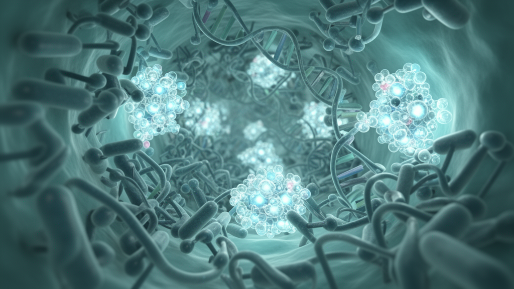
“I think of it as a city,” Dobbs explained. “The molecular machines I work on are like little factories that produce things, while some of my colleagues have been studying systems that transport and process those products. Together, they form different parts of a living molecular community inside the cell – much like a city with different specialised activities all working together.” It is precisely this focus on interactions between molecular machines, rather than on the machines in isolation, that distinguishes the two papers from the bulk of prior structural biology and marks them as proofs of concept for a new analytical paradigm.
The second paper, led by Rasmus Jensen of the Mahamid group, approached the cell from the opposite direction. Rather than starting with a specific biological question, Jensen began with an observation: something unusual and unexplained was visible in the tomographic images of the cell. “Joe started with a biological question he wanted to answer,” Jensen said. “I started from the opposite direction. We’d seen something interesting in the cell and worked backwards to figure out what it was, eventually discovering that it’s a new configuration of a molecular machine that helps proteins either be exported out of the cell or inserted into its membrane.” That reverse-engineering approach led the team to a discovery that no one had anticipated: an entirely new protein-folding machinery operating at the cell surface.
By combining cryo-ET imaging with proteomics—the large-scale identification of the proteins present in a sample—and computational protein structure prediction, the researchers determined that this newly characterized machinery contains the well-known Sec-translocon, a protein channel embedded in the cell membrane that functions as a doorway through which newly made proteins exit the cell or become inserted into the membrane itself. Alongside the translocon, the team identified three previously unknown proteins that assist transported proteins in folding into their correct functional shapes after they emerge at the cell surface. The cryo-ET data yielded the most detailed images to date of the conserved portion of the bacterial protein transport machinery, showing how proteins fresh from the ribosome begin their journey through the membrane and revealing that transport is closely linked to a folding system operating outside the cell—a location where such machinery had not been described before in this organism.
“Working together with many colleagues at EMBL, we combined expertise in microbiology, proteomics, bioinformatics, structural biology, and integrative modelling,” Mahamid said. “This paper tells the entire story of the discovery process: visualising a new cellular machinery, identifying the genes and proteins, and figuring out how it all functions. That’s a remarkable achievement and opens new ways to identify new complexes and functions in different systems.” The workflow demonstrated in the study—imaging an unknown structure, tracing it back to its genetic basis, and then characterizing its function—offers a template that other laboratories can apply to organisms far more complex than a minimal bacterium.
Indeed, the broader significance of both studies lies less in any single finding than in the conceptual foundation they establish. “In both Joe’s work and mine, we’ve started with what we’d call a minimal cell where everything is relatively simple,” Jensen noted. “We’ve advanced what is conceptually possible in this minimal bacterium and established ways to apply these methods to more complex organisms.” Mahamid frames the future in similar terms: “The future is studying molecular communities. Decades of structural biology done on isolated molecules provided us with detailed views of their structures and functions. In the past, we just didn’t have the technology to study their more complex interactions and cross-talk. This work points again to how cryo-ET is an enabling technology, allowing us to see new things and connectivity inside cells. Its ability to show us more seems only limited by our asking the right questions.” As cryo-ET continues to mature, the molecular cities inside cells—bacterial and human alike—are finally beginning to reveal not just their buildings, but the bustling traffic that connects them.
Subject of Research: Visualizing interconnected protein production, transport, and folding machineries in intact bacterial cells using cryo-electron tomography
Article Title: The bustling molecular communities inside cells
Article References: The bustling molecular communities inside cells. (n.d.). Original publication
Image Credits: AI Generated
DOI: Not provided
Keywords: cryo-electron tomography, Mycoplasma pneumoniae, ribosomes, transcription, translation, Sec-translocon, protein folding, protein transport, structural biology, supercomplexes, cell membrane, EMBL
News Source: Morgan Morrow. (October 18, 2026). Inside the Molecular Cities of Bacteria: New Imaging Reveals How Cellular Factories Work Together. Scienmag.