
A groundbreaking advancement in the preoperative marking of peripheral pulmonary lesions (PPLs) promises to revolutionize thoracic surgery precision and patient outcomes. Pulmonologist and interventional pulmonology expert Dr. Ganesh Krishna of Sutter’s Palo Alto Medical Foundation and El Camino Hospital has spearheaded a multisite retrospective study exploring the efficacy of a novel technique involving indocyanine green-soaked fiducial (ICG-F) markers. This new approach aims to enhance the visualization and accurate localization of lung nodules prior to surgical resection, addressing a critical challenge in minimally invasive thoracic procedures.
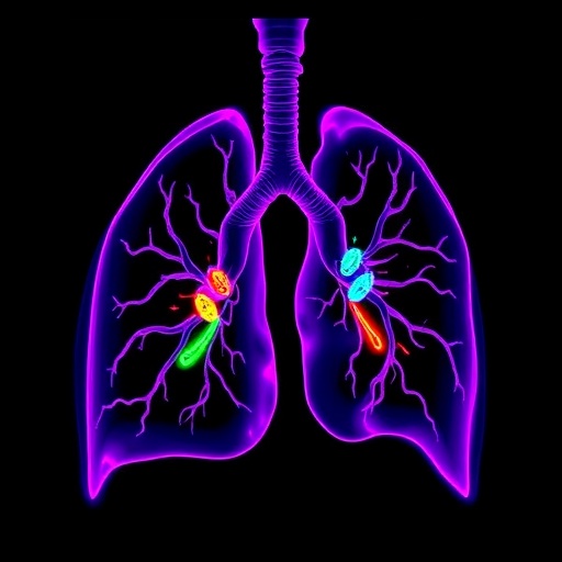
Peripheral pulmonary lesions, often small and difficult to detect during surgery, pose significant challenges for surgeons aiming for lung-sparing resections. Traditional marking methods can be invasive, less accurate, or logistically complicated, particularly when coordination between bronchoscopy and surgery under a single anesthetic event proves difficult. Dr. Krishna’s study sought to overcome these barriers by utilizing bronchoscopic delivery of ICG-F markers, a fluorescent dye combined with fiducial markers that can be visualized intraoperatively through near-infrared imaging.
In this retrospective analysis, Dr. Krishna and collaborators collected data from four medical centers: El Camino Hospital/Palo Alto Medical Foundation, Advocate Aurora Medical Center of Kenosha, Emory University, and the University of California at Davis. The study compared outcomes of 54 PPLs localized using ICG-F markers against 63 unmarked lesions. Crucially, the decision to utilize this technique was left to the discretion of local medical teams, allowing a pragmatic assessment of its real-world applicability.
The findings demonstrated that all ICG-F marked lesions were distinctly visible during surgery either immediately following the bronchoscopy or up to 13 days later, a remarkable duration that adds logistical flexibility previously unavailable. Analysis revealed that the ICG-F marked nodules were smaller on axial and coronal measurements compared to unmarked ones, suggesting that the new technique supports high-precision localization suitable for lung-sparing operations that aim to preserve maximum healthy tissue.
Interestingly, despite the smaller specimen weight and size in the ICG-F group, surgical procedures required significantly longer operative times—on average, an additional 29 minutes. Dr. Krishna attributes this increase not to inefficiency but rather the meticulous dissection required to excise complex, deeper anatomical structures during segmentectomies or lobectomies facilitated by enhanced lesion localization. This precision, while time-consuming, may translate into better oncologic outcomes and reduced postoperative morbidity.
The study emphasizes the safety and accuracy of bronchoscopic ICG-F marking as a minimally invasive alternative to conventional methods, potentially widening access to thoracic surgery in centers lacking readily available specialized surgeons. The technique also addresses logistical hurdles by decoupling the marking procedure from immediate surgery, a significant advantage in busy healthcare settings aiming to optimize resource allocation.
However, as with many pioneering studies, limitations exist. The retrospective and non-randomized design introduces the possibility of selection bias and confounding variables that a prospective, randomized controlled trial would better mitigate. Additionally, the learning curve associated with employing novel imaging and marking technologies must be acknowledged, underscoring the need for comprehensive training and standardization as adoption expands.
Early and accurate identification of malignant lung nodules is critical, as lung cancer remains the leading cause of cancer-related deaths in the United States, with survival rates heavily dependent on disease stage at diagnosis. Innovations such as the ICG-F marking technique directly contribute to the evolving paradigm of early intervention, where improved surgical precision equates to increased rates of cure and lung function preservation.
Indocyanine green (ICG) has been used previously in various medical imaging applications due to its fluorescence properties under near-infrared light. However, combining it with fiducial markers and bronchoscopic delivery represents a significant leap forward. This allows surgeons not only to locate lesions visually but also to navigate anatomical complexities with real-time feedback—enhancing confidence in minimal resections.
Moreover, the prospect of marking PPLs days before surgery offers important implications for patient scheduling and resource management. This can reduce the burden on anesthetic services and potentially lower patient exposure to risks associated with prolonged operative anesthesia. Such operational efficiencies could improve patient throughput and satisfaction within healthcare systems.
Dr. Krishna and his team envision further research to evaluate long-term patient outcomes, including post-surgical lung function, recurrence rates, and quality of life metrics. Integration of this technology with emerging robotic-assisted thoracic surgery platforms could also amplify its impact, enabling surgeons to harness precision imaging alongside enhanced dexterity.
In conclusion, the introduction of bronchoscopic indocyanine green-soaked fiducial markers heralds a promising advancement in thoracic surgery, potentially shifting standards toward safer, more precise, and accessible treatment for peripheral pulmonary lesions. While further validation through prospective studies is needed, this technique embodies the ongoing convergence of innovative imaging, minimally invasive procedures, and patient-centric care in pulmonary medicine.
Subject of Research: Novel bronchoscopic technique using indocyanine green-soaked fiducial markers for localization of peripheral pulmonary lesions prior to surgical resection.
Article Title: Not specified in the provided content.
News Publication Date: Not specified in the provided content.
Web References:
CHEST® Pulmonary Journal: https://www.chestpulmonary.org/article/S2949-7892(24)00097-7/fulltext
Lung Cancer Statistics (American Cancer Society): https://www.cancer.org/cancer/lung-cancer/about/key-statistics.html#:~:text=Lung%20cancer%20is%20by%20far,breast%2C%20and%20prostate%20cancers%20combined.
Sutter Health Annual Report: https://www.sutterhealth.org/annualreport-2023
References: Retrospective multisite study led by Dr. Ganesh Krishna published in CHEST Pulmonary.
Keywords: Surgery, Pulmonary Lesions, Interventional Pulmonology, Thoracic Surgery, Indocyanine Green, Fiducial Markers, Lung Cancer, Bronchoscopy, Minimally Invasive Surgery.
Tags: bronchoscopic delivery methodsDr. Ganesh Krishna researchenhancing surgical precisionfiducial markers in surgeryindocyanine green dye technologyinnovative surgical techniquesminimally invasive thoracic surgerymultisite retrospective study in medicinenear-infrared imaging in surgeryperipheral pulmonary lesionspreoperative marking for lung lesionspulmonary nodule localization
