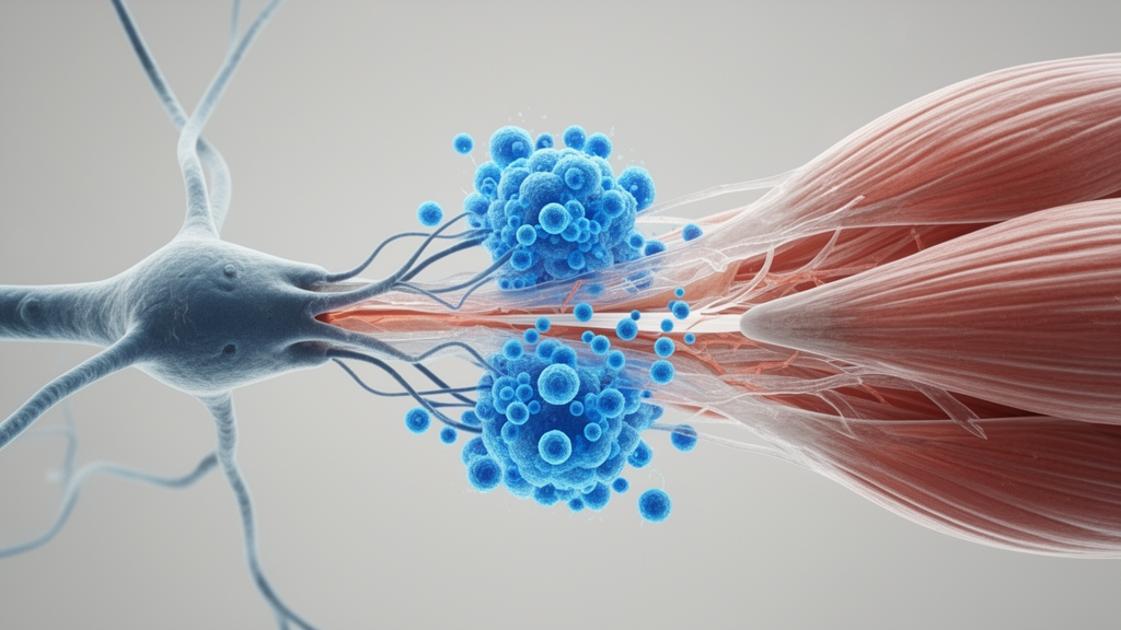
A comprehensive scientific review published in Frontiers in Physiology reveals that the progressive deterioration of skeletal muscle mass and strength defining sarcopenia is primarily initiated by the inflammatory remodeling of the local immune microenvironment at the neuromuscular junction rather than starting as passive muscle fiber wasting. Synthesizing evidence across immunology and neuromuscular physiology, the researchers propose that structural disintegration of this vital chemical synaptic bridge occurs significantly earlier along the pathological timeline than detectable motor neuron loss or muscle fiber atrophy. Advancing age is characterized by chronic, low-grade systemic inflammation—termed inflammaging—which permeates skeletal muscle tissue and reshapes the resident immune population. In older individuals carrying a heightened inflammatory burden, local immune cells undergo profound phenotypic reprogramming, transitioning from supportive homeostatic maintainers into active instigators of synaptic injury. This shift exploits the unique intrinsic vulnerabilities of the junction, including an absence of a blood-nerve barrier and extreme sensitivity to oxidative stress, thereby initiating an autonomous pathological cascade that precedes overt muscular decay.
The clinical significance of this newly proposed paradigm addresses a persistent therapeutic hurdle in geriatric medicine since the World Health Organization formally classified sarcopenia as an independent clinical disease entity in 2016. Despite escalating prevalence and severe downstream consequences—ranging from loss of physical independence and heightened fall frequencies to elevated socioeconomic burdens and increased mortality—targeted therapies capable of reliably reversing functional loss remain largely unavailable. Current primary interventions relying on resistance exercise protocols combined with structured nutritional supplementation frequently encounter widespread intervention resistance, while emerging pharmacological strategies such as exercise mimetics, nicotinamide adenine dinucleotide precursors, and myokine-directed monoclonal antibodies have demonstrated limited efficacy in halting progression. The authors argue that this therapeutic gap stems directly from traditional research paradigms that disproportionately focused on intrinsic muscle defects, such as muscle satellite cell exhaustion and myofiber mitochondrial dysfunction, while neglecting the specialized neuro-immune-muscular signaling interface that governs excitation-contraction coupling and preserves motor endplate stability.
Pathological and morphological evaluations of the aging neuromuscular junction demonstrate unmistakable structural aberrations that emerge well before the onset of measurable myofiber atrophy. Postsynaptic acetylcholine receptor clusters, which normally form dense and continuous specialized plaques, fracture into fragmented islands and develop abnormal micro-perforations alongside flattened junctional folds and thickened basal laminae. Simultaneously, presynaptic motor axon terminals retract, reducing total synaptic contact area, while swollen nerve endings packed with abnormal mitochondria coexist with aberrant, inefficient regenerative axon sprouts. This ongoing structural destabilization increases the proportion of partially denervated endplates across aging muscle tissue, reflecting a scenario where primary terminal function falters faster than compensatory reinnervation can occur. Furthermore, because healthy neuromuscular transmission operates with a generous physiological safety factor, significant structural deterioration can quietly accumulate without causing immediate muscle failure. Consequently, aging synapses frequently transition into a silent, sub-functional state long before overt electrical transmission failure occurs, causing conventional functional testing to systematically underestimate early disease development.
The inherent susceptibility of the neuromuscular junction to immune-mediated destruction is intrinsically tied to its extraordinary metabolic demands and unique microanatomical exposure. Neurotransmission requires continuous, ATP-dependent vesicle recycling, transmitter synthesis, and quantal release of acetylcholine, rendering the presynaptic nerve terminal one of the most mitochondria-dense subcellular regions within the entire human body. As tissue ages, the vital quality control pathway governed by PINK1 and Parkin-mediated mitophagy progressively deteriorates, causing damaged mitochondria, misfolded structural proteins, and mutated mitochondrial DNA to accumulate within the confined synaptic space. Postsynaptic mitochondrial failure further impedes local cellular energetics, delaying axonal reinnervation and weakening the agrin, LRP4, MuSK, and rapsyn signaling axis essential for anchoring receptor assemblies. Crucially, the peripheral synaptic cleft entirely lacks a protective blood-nerve barrier, directly exposing vulnerable nerve terminals, receptor arrays, and specialized extracellular matrix components to circulating cytokines, matrix metalloproteinases, and classical complement proteins such as C1q that are otherwise excluded from protected nervous system compartments.
Within the aging skeletal muscle stroma, cellular immunity undergoes extensive remodeling characterized by the emergence of specialized, disease-associated myeloid and lymphoid subsets. In healthy young muscle, tissue-resident macrophages secrete pro-regenerative molecules such as insulin-like growth factor-1 that foster an anti-inflammatory microenvironment and direct muscle satellite cell proliferation during repair. In aging tissue, leaked mitochondrial DNA triggers toll-like receptors and the NLRP3 inflammasome, skewing macrophages toward a persistent senescence-associated secretory phenotype. Rather than conforming to neat binary classifications of classical pro-inflammatory M1 or reparative M2 states, aged macrophages occupy complex, intermediate phenotypes; single-cell transcriptomics reveal an enrichment of TREM2-expressing lipid-associated macrophages that robustly transcribe catabolic mediators, including Secreted Phosphoprotein 1. Concurrently, injured muscle fibers release extracellular vesicles carrying BAG6 that bind to NCR3 receptors on tissue-resident T cells, promoting local T-helper 17 polarization. These Th17 cells upregulate surface CD6 expression, further amplifying Secreted Phosphoprotein 1 synthesis while local loss of interleukin-33 production impairs regulatory T cell recruitment.
Once shifted into a destructive profile, immune cells unleash direct molecular and physical assaults against the physical scaffold of the neuromuscular junction. Uncontrolled local macrophages engage in aberrant synaptic pruning, engulfing partially intact presynaptic terminals and postsynaptic membrane structures while simultaneously secreting destructive proteolytic enzymes, notably matrix metalloproteinase-14 and elastase. These proteases directly dismantle the specialized synaptic basal lamina, degrading vital scaffolding molecules such as agrin, laminin, and type IV collagen that physically anchor acetylcholine receptor clusters and stabilize junctional folds. In parallel, persistently elevated local concentrations of tumor necrosis factor-alpha and interleukin-6 traverse the compromised basement membrane to engage postsynaptic sarcolemmal receptors. This engagement activates the nuclear factor-kappa B intracellular signaling cascade, inducing rapid nuclear translocation that upregulates muscle-specific E3 ubiquitin ligases, specifically MuRF1 and Atrogin-1. This transcriptional activation not only accelerates myofibrillar protein breakdown but also actively represses the gene networks responsible for maintaining receptor density, substantially degrading the electrical safety factor.
The breakdown of neuromuscular integrity is exacerbated by the failure of the non-neuronal cholinergic system, an endogenous anti-inflammatory axis that normally restrains microenvironmental immune reactivity. Under physiological circumstances, activated T and B lymphocytes express choline acetyltransferase, allowing them to synthesize and secrete non-neuronal acetylcholine in response to sympathetic nerve signals and specific cytokine cues. This locally derived acetylcholine binds to alpha-7 nicotinic acetylcholine receptors located on the surface of surrounding macrophages, triggering a biochemical cascade that suppresses the synthesis of damaging inflammatory cytokines. In sarcopenic muscle, down-regulated choline acetyltransferase expression combined with altered lymphocyte subsets causes this protective cholinergic anti-inflammatory reflex to collapse. Lacking negative feedback restraint, uninhibited macrophages and mast cells undergo sustained respiratory bursts, releasing overwhelming surges of reactive oxygen and nitrogen species. These oxidants attack adjacent presynaptic mitochondria, collapsing membrane potentials, exhausting ATP production, impairing vesicle mobilization, and opening the mitochondrial permeability transition pore to release cytochrome c, thereby triggering terminal apoptotic degeneration.
A critical conceptual contribution of the review is the formal delineation of low-affinity, autoimmune-like attacks directed against endogenous synaptic structures during advanced aging. Driven by persistent local antigen exposure and molecular mimicry, infiltrating T helper cells facilitate the production of low-affinity autoantibodies by B cells against acetylcholine receptors and synaptic organizers like MuSK and LRP4. These autoantibodies activate the classical complement pathway, depositing membrane attack complexes directly onto postsynaptic folds and accelerating enzymatic membrane lysis. The authors emphasize that this phenomenon differs fundamentally from classical myasthenia gravis, as it does not rely on robust germinal center formation or high-affinity memory clones, but rather represents steady-state leakage of antibodies against an already weakened target. In young individuals, healthy neuromuscular junctions easily tolerate low-level antibody deposition thanks to abundant functional reserves; in contrast, the aged junction, compromised by mitochondrial fatigue and degraded receptor density, suffers catastrophic electrical transmission failure even when confronted with minimal immunological assault.
Acting as specialized resident sentinels, terminal Schwann cells serve as the decisive cellular link between initial synaptic distress and persistent inflammatory cell recruitment. These non-myelinating glial cells normally envelop motor nerve endings, sensing neurotransmission fidelity, directing synaptic maintenance, and producing trophic factors such as transforming growth factor-beta-1. However, under progressive energetic failure and hypothalamic Sirt1 signaling decline, terminal Schwann cells undergo functional degeneration and switch from protective guardians into active drivers of inflammation. Stressed terminal Schwann cells dramatically upregulate chemokine ligands, including CCL2 and CCL12, generating an intense chemotactic gradient that recruits circulating monocytes, neutrophils, and adaptive lymphocytes into the perisynaptic space. In the aged, hypoxic microenvironment, these recruited cells cannot transition toward regenerative phenotypes and instead remain locked in pro-inflammatory states. They continuously secrete tumor necrosis factor-alpha and interleukin-6 while blocking restorative interleukin-10 and transforming growth factor-beta pathways, creating an enduring vicious cycle wherein ongoing synaptic breakdown releases damage-associated molecular patterns that summon fresh waves of destructive immune cells.
Translating the neuro-immune-muscular paradigm into clinical practice requires moving beyond simple measurements of gross muscle bulk toward sensitive functional evaluations of the synaptic apparatus. Diagnostic protocols must incorporate electrophysiological modalities such as single-fiber electromyography to quantify neuromuscular jitter and impulse blocking, alongside surface electromyographic motor unit number estimation to identify subclinical denervation before irreversible fibroadipogenic degeneration settles into contractile tissue. Therapeutic strategies must likewise evolve from focusing strictly on muscle fibers toward targeted modulation of the synaptic microenvironment. Promising avenues include lifestyle interventions and dietary regimens that preserve regulatory T cell competence, pharmacological reprogramming of myeloid cells via tyrosine kinase inhibitors or PPAR-gamma agonists, and cautiously applied complement or FcRn inhibitors tailored specifically to frail patients exhibiting documented synaptic autoimmunity. By targeting the immune-synaptic axis during the critical window before structural collapse becomes permanent, clinicians may finally establish effective disease-modifying therapies capable of halting or reversing the debilitating progression of sarcopenia.
Subject of Research: Immune microenvironment remodeling and neuromuscular junction degeneration in sarcopenia
Article Title: Inflammaging at the neuromuscular junction: immune microenvironment remodeling as the trigger for sarcopenia
Article References: Sun, X., Nie, J., Cai, X., Ma, X., & Miao, Y. (2026). Inflammaging at the neuromuscular junction: immune microenvironment remodeling as the trigger for sarcopenia. Frontiers in Physiology, 17, Article 1988379. https://doi.org/10.3389/fphys.2026.1988379
Image Credits: AI Generated
DOI: 10.3389/fphys.2026.1988379
Keywords: neuromuscular junction, sarcopenia, inflammaging, terminal Schwann cells, acetylcholine receptors, synaptic pruning, non-neuronal cholinergic system, muscle satellite cells, mitochondrial dysfunction, TREM2 macrophages, complement C1q, single-fiber electromyography, MuSK, LRP4, pro-inflammatory cytokines, motor endplate denervation, Secreted Phosphoprotein 1, choline acetyltransferase
News Source: Drew Townsend. (November 17, 2026). Immune Microenvironment Breakdown at Neuromuscular Junctions Drives Early Sarcopenia. Scienmag.