


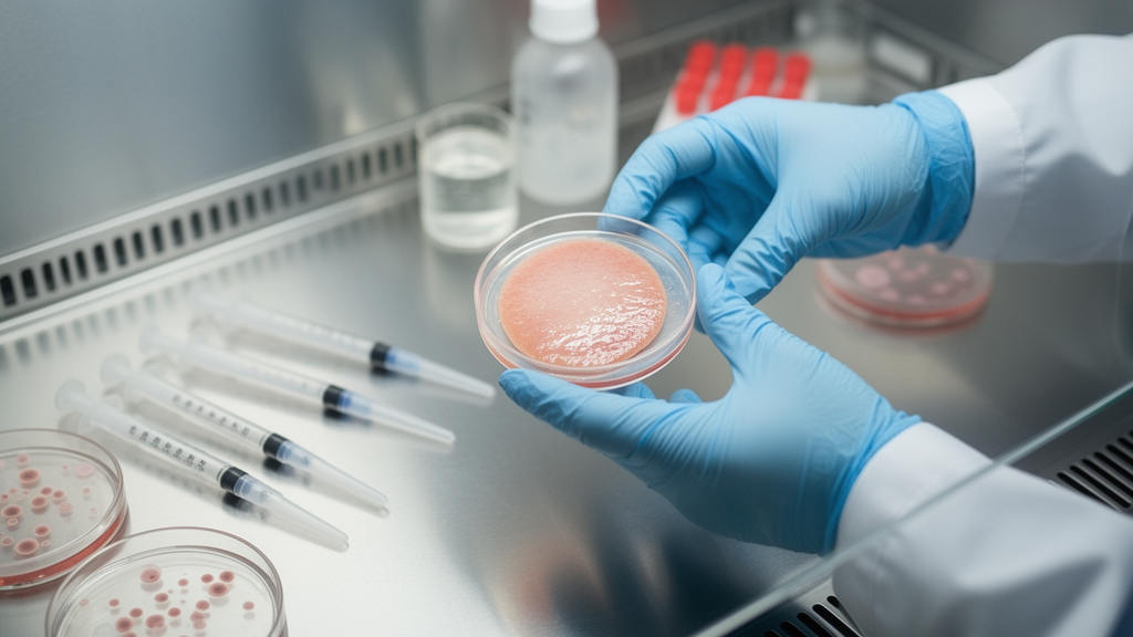
Researchers in Brazil have turned surplus skin from abdominoplasty surgeries into a working laboratory model for Staphylococcus aureus infections, keeping the tissue alive and structurally intact long enough to track bacterial growth and antibiotic response for up to seven days. The pilot study, published in International Microbiology, standardized the human organotypic skin explant culture, or hOSEC, as an alternative to animal skins and simplified cell cultures for testing how this common pathogen colonizes human skin and how antimicrobials push back.
Because the tissue would otherwise be discarded as surgical waste, the approach aligns with the 3Rs principles of reduction, refinement, and replacement in animal research. The researchers cut 8-millimeter and 10-millimeter punch biopsies from the samples and placed them dermal-side down on filter paper over metal grids in culture plates, keeping the epidermis exposed at the air-medium interface.
Before any bacteria entered the picture, the team had to confirm the explants stayed alive. A tetrazolium chloride assay, which measures mitochondrial activity, showed the tissue weathered the processing remarkably well. After four hours of antibiotic decontamination at 2 to 8 degrees Celsius, viability measured at 3 hours post-decontamination showed a median of 64 percent, but after 24 hours in culture it reached a median of 125.5 percent of baseline, and after five days it remained near baseline at a median of 105.5 percent. When the researchers applied a stronger antibiotic treatment directly, viability held at 97 percent, a drop of only 3 percent. Histology of uninfected explants confirmed the full architecture of human skin: a keratinized stratified squamous epidermis, an intact dermal-epidermal junction, organized collagen bundles, fibroblasts, resident immune cells, blood vessels, sweat glands, hair follicles, and even the hypodermis.
Three infection protocols were tested. In the first, bacteria were injected directly into the dermis to mimic deep tissue infection. In the second, S. aureus was applied topically to intact epidermis. In the third, a 4-millimeter wound was punched into the explant before topical inoculation, simulating an infected wound. All assays used 10 microliters of inoculum at a concentration of 10⁶ CFU/mL, and the strains included a susceptible reference isolate (ATCC 25923), a resistant isolate (ATCC BAA-1026), and a methicillin-resistant strain (NCTC 12493).
The intradermal model delivered a dramatic result. Untreated explants injected with the MRSA strain proliferated beyond 1 × 10⁸ CFU/mL within 24 hours, while antibiotic-treated tissue suppressed growth to an average of 2 × 10² CFU/mL, roughly a 99.9 percent reduction. The antibiotic added to the culture medium was absorbed through the dermis, controlling bacterial growth in place. The researchers noted, however, that bacteria leaked into the surrounding medium, so this model does not confine the infection to a controlled site.
Topical application to intact skin proved less durable. Counts started above 1 × 10⁸ CFU/mL but fell sharply by 12 and 24 hours in both treated and saline control groups, leaving too little recoverable bacteria to sustain a colonization curve. That limitation pushed the team toward the wound model, which became the centerpiece of the study.
In the wound model with the resistant strain, saline-treated explants maintained high counts above 1 × 10⁸ CFU/mL for the first six hours. Antibiotic treatment cut counts significantly by 3 hours (median 190 versus 1000 CFU per 10 microliters, p = 0.0045) and further by 6 hours (median 62 versus 1000, p = 0.0061) and 12 hours (median 11 versus 395.5, p < 0.0001), though both groups still carried mean counts of 1.8 × 10⁷ and 9.6 × 10⁷ CFU/mL at 3 hours and 1.8 × 10⁶ and 4.2 × 10⁷ CFU/mL at 12 hours. By 24 hours, saline-treated tissue still carried a mean of 2.7 × 10⁶ CFU/mL, and antibiotic-treated tissue 1.7 × 10⁶ CFU/mL. Viable bacteria persisted in the explants for the full observation period: at 72, 120, and 168 hours, antibiotic-treated fragments yielded 4 × 10², 1.5 × 10², and 2.5 × 10² CFU/mL respectively, while saline-treated fragments yielded 3.2 × 10³, 1 × 10², and 0 CFU/mL. The persistence of CFU in antibiotic-treated fragments after seven days suggests survival of resistant subpopulations, a pattern the authors link to phenotypic heterogeneity and selection under antimicrobial pressure.
Histopathology revealed the cost of colonization to the tissue itself. Beginning at 12 hours, infected explants showed apoptotic keratinocytes and early desquamation of the stratum corneum. After 24 hours, necrosis appeared in the basal layer alongside spreading apoptosis. By day three, keratinocyte death extended through the stratum granulosum and stratum spinosum, and collagen fibers in the dermis became disorganized. At seven days, the infected tissue displayed intraepidermal acantholysis, loss of the dermal-epidermal junction, and nuclear changes consistent with cell death, both at the wound edge and in distal regions. Basophilic granular structures suggestive of bacterial aggregates, compatible with biofilm formation, were visible in the wound area under high magnification.
The authors position hOSEC against commercial platforms such as Genoskin’s WoundSkin, which they note can be costly and subject to import restrictions in Brazil. Their model, built from locally sourced surgical waste, offers a cost-effective alternative that preserves native tissue complexity. Compared with 3D cultures, spheroids, and organoids, the explant retains epidermal stratification, extracellular matrix, and the interplay of keratinocytes, fibroblasts, and resident immune cells. The researchers acknowledge limitations: the explants lack blood flow and a resident microbiota, bacterial recovery can be confounded by intracellular persistence or small colony variants, and residual antibiotic from decontamination cannot be fully excluded. As a pilot study using explants from six patients, the findings establish feasibility rather than definitive performance. Even so, the work demonstrates that living human skin explants, obtained from surgical waste, can sustain measurable bacterial colonization and antibiotic response for up to seven days, supporting the model's use for ex vivo evaluation of antimicrobial penetration, local efficacy, and bacterial dynamics in human skin–mimetic conditions.
Subject of Research: An ex vivo human skin explant model for studying Staphylococcus aureus colonization and antibiotic testing
Article Title: Human organotypic skin explant culture (hOSEC): an alternative approach to Staphylococcus aureus colonization assays
Article References: Lisboa, L. F. T., Barbosa, L. D., Giarola, F., Galani, L. C., da Costa, W. P., Frade, M. A. C., Monteiro, M. C., & Lima, F. R. (2026). Human organotypic skin explant culture (hOSEC): an alternative approach to Staphylococcus aureus colonization assays. International Microbiology. https://doi.org/10.1007/s10123-026-00908-y
Image Credits: AI Generated
DOI: 10.1007/s10123-026-00908-y
Keywords: Staphylococcus aureus, skin explant culture, hOSEC, antimicrobial testing, MRSA, wound infection, tissue viability, histopathology, ex vivo model, 3Rs, antibiotic resistance, skin colonization
News Source: Drew Townsend. (October 30, 2026). Human organotypic skin explant culture: an ex vivo model for Staphylococcus aureus colonization and antimicrobial testing. Scienmag.



