Uterine fibroids are among the most common tumors in women of reproductive age, benign growths of the muscular wall of the uterus that can cause heavy menstrual bleeding, pelvic pressure, pain, and a level of daily disruption that many patients describe as life-altering. For decades, the standard answers were hormonal therapy, surgical myomectomy, or hysterectomy, each with its own trade-offs in recovery time, fertility implications, and surgical risk. Over the past two decades, a radically different option has moved from experimental curiosity to mainstream clinical practice: high-intensity focused ultrasound, or HIFU, a technique that destroys fibroid tissue from outside the body using concentrated sound waves, with no incision at all. Now a new retrospective cohort study from Changde First People’s Hospital in Hunan Province, China, published in BMC Cancer, has tackled one of the most consequential unanswered questions about this technology: how much of the fibroid actually needs to be destroyed for patients to feel meaningfully better?
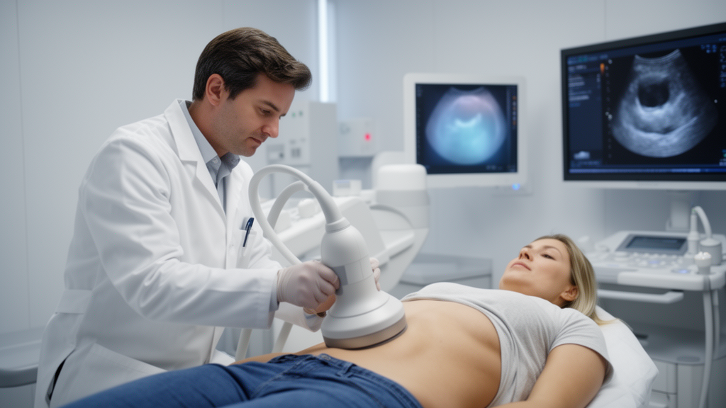
The technical logic behind HIFU is elegant. An extracorporeal transducer focuses multiple ultrasound beams on a target inside the fibroid, raising the temperature at the focal point high enough to induce coagulative necrosis, the irreversible thermal death of tissue, while sparing the skin, bowel, and surrounding structures along the beam path. Because the treated tissue is killed in place rather than removed, the critical question after every session is not how much tissue was excised, as it would be in surgery, but how much tissue was actually devascularized. Contrast-enhanced magnetic resonance imaging or ultrasound provides the answer in the form of the non-perfused volume ratio, or NPVR, the proportion of the fibroid volume that no longer takes up contrast agent, indicating tissue whose blood supply has been permanently interrupted. A high NPVR means the ablation covered most of the fibroid; a low NPVR means substantial viable tissue remains behind.
What the Changde team, led by Lei Dong and corresponding author Yuxin Zhou, did was to ask whether the achieved NPVR, measured immediately after treatment, actually predicts how patients fare months later. The study included 168 women with symptomatic uterine fibroids who underwent HIFU and completed six months of follow-up. For descriptive purposes, patients were sorted into three groups according to their NPVR: below 80 percent, 80 to below 90 percent, and 90 percent or above. But the analysis went far beyond simple group comparisons. The investigators used linear mixed-effects models to track longitudinal changes in symptom severity and health-related quality of life, and multivariable regression to isolate the independent contribution of NPVR after adjusting for baseline symptom scores, fibroid anatomical characteristics such as size and location, and other prespecified covariates. This statistical architecture matters, because it separates the effect of how thoroughly the fibroid was ablated from the effect of how severe the fibroid was to begin with.
The headline finding is strikingly consistent: higher achieved NPVR was associated with greater fibroid volume reduction and more favorable symptom and quality-of-life outcomes throughout follow-up. In the continuous-NPVR longitudinal analysis, the time-by-NPVR interaction was highly significant for both the Symptom Severity Scale, with F equal to 32.953 and P below 0.001, and for health-related quality of life, with F equal to 19.324 and P below 0.001. In plain terms, patients whose fibroids were more completely ablated did not merely start from a better position; their trajectories diverged over time, with the gap between high-NPVR and low-NPVR patients widening as the months passed. This is the kind of signal that clinicians look for when deciding whether an imaging marker is merely decorative or genuinely informative about clinical destiny.
The adjusted six-month analyses reinforce the same conclusion with impressive precision. After accounting for baseline outcome measures and fibroid anatomy, each increment in NPVR remained independently associated with a lower six-month Symptom Severity Scale score, with a standardized beta of minus 0.426 and a 95 percent confidence interval spanning minus 0.547 to minus 0.305, and with a higher six-month quality-of-life score, with a beta of 0.356 and a confidence interval of 0.241 to 0.471, both P below 0.001. The Symptom Severity Scale is part of the validated Uterine Fibroid Symptom and Quality of Life questionnaire, a widely used instrument in which scores of 20 points or more are considered clinically meaningful. At six months, 119 of the 168 patients, or 70.83 percent, achieved the prespecified clinical-response criterion of an SSS reduction of at least 20 points. Higher NPVR was associated with greater odds of achieving this response, with an odds ratio of 1.102 per unit increment and a confidence interval of 1.059 to 1.150.
For readers wondering whether NPVR alone can tell a clinician who will respond and who will not, the exploratory receiver operating characteristic analysis offers a sobering technical answer. The area under the curve was 0.745, with a confidence interval of 0.659 to 0.831, which the authors characterize as moderate discrimination. In practical terms, NPVR is a strong post-treatment indicator, but it is not a crystal ball. A patient with a high NPVR is more likely to respond, but the marker cannot substitute for the full clinical picture, including fibroid anatomy, baseline symptom burden, and individual healing dynamics. This nuance is important for preventing the kind of oversimplification that often accompanies viral coverage of medical imaging markers, where a single number is presented as destiny rather than as one informative variable among many.
Why does this matter so much for the future of fibroid care? HIFU is already attractive because it avoids incisions, general anesthesia in many protocols, and the prolonged recovery associated with surgery. But its Achilles heel has always been variability in ablation extent. Fibroids differ enormously in their acoustic properties, their vascularity, their distance from the skin, and their proximity to bowel or the sacrum, and operators must balance the desire for complete ablation against the risk of thermal injury to surrounding structures. The new study suggests that the achieved NPVR is not just a technical scorecard for the operator; it is a genuine predictor of how much symptom relief and quality-of-life benefit the patient will experience. That transforms NPVR from an imaging curiosity into a piece of information that can guide post-treatment counseling, follow-up intensity, and decisions about whether additional sessions or adjunctive therapies are warranted.
At the same time, the authors are emphatic about the limits of their findings, and their caution deserves equal billing with the positive results. This was a single-center retrospective cohort study, not a randomized trial, and the association between NPVR and outcomes is observational. The statistical adjustments controlled for baseline measures and anatomy, but retrospective designs cannot exclude residual confounding, and the results reflect the practice patterns and patient population of one institution. Most importantly, the investigators warn explicitly that NPVR should not be interpreted as a therapeutic target to be maximized irrespective of procedural safety. Pushing ablation extent higher can increase the risk of thermal injury to skin, bowel, nerves, or the endometrium, and the optimal balance between ablation completeness and safety must be determined in prospective studies rather than inferred from retrospective associations.
There is also a broader scientific context that makes this study more than a local quality-improvement exercise. Uterine fibroids affect a large proportion of women during their reproductive years, and the search for less invasive, fertility-preserving treatments has become one of the most active frontiers in gynecology. HIFU sits at the intersection of physics, engineering, and clinical medicine, requiring precise acoustic targeting, real-time monitoring, and careful thermal dosimetry. Studies like this one, which connect the physics of ablation to validated patient-reported outcomes, are exactly the kind of translational evidence needed to move the field from proof of concept toward personalized treatment planning. If future prospective research confirms these findings, NPVR could become a standard component of post-HIFU assessment, informing both clinical follow-up and shared decision-making between patients and physicians.
For now, the message for patients and clinicians is measured but genuinely encouraging. The Changde study provides some of the clearest retrospective evidence to date that how thoroughly a fibroid is ablated with focused ultrasound is associated with how much better patients feel and how much their quality of life improves over the following six months. It does not promise that every patient will respond, and it does not license operators to chase higher NPVR at any cost. But it does give the field a measurable, imaging-based variable that tracks with clinically meaningful benefit, and it sets the stage for the prospective trials that will determine whether NPVR can be safely optimized as part of routine fibroid care. In a specialty where the goal is increasingly to destroy disease without ever touching the body with a scalpel, that is a milestone worth noting.
Subject of Research: uterine fibroids
Article Title: Association between HIFU ablation extent and symptom relief and quality of life benefits in uterine fibroids: a retrospective analysis based on imaging and clinical indicators
Article References: Dong, L., Wang, L., Zeng, Y., Fu, W., & Zhou, Y. (2026). Association between HIFU ablation extent and symptom relief and quality of life benefits in uterine fibroids: a retrospective analysis based on imaging and clinical indicators. BMC Cancer. https://doi.org/10.1186/s12885-026-16934-y
Image Credits: AI Generated
DOI: 10.1186/s12885-026-16934-y
Keywords: uterine fibroids, HIFU, non-perfused volume ratio, minimally invasive surgery, quality of life, medical imaging, Association, between, ablation, extent, symptom, relief
News Source: Ophelia Keating. (October 17, 2026). How Much Fibroid Tissue Doctors Destroy With Sound Waves Predicts Who Feels Better. Scienmag.