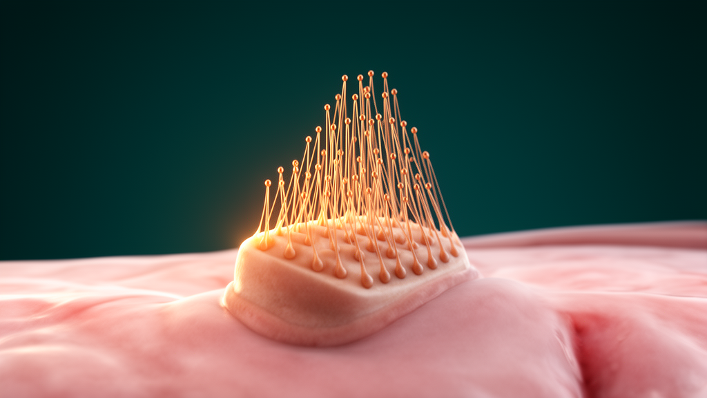
Stereocilia, the tiny actin-filled protrusions that crown inner-ear hair cells and form staircase-like bundles essential for hearing, arise through a carefully choreographed sequence of changes on the cells’ apical surface, according to a new study of the developing mouse cochlea published in Frontiers in Cell and Developmental Biology.
Researchers Suraj Ranganath Chakravarthy and Raj Ladher of the Tata Institute of Fundamental Research National Centre for Biological Sciences in Bengaluru, together with Thomas S. van Zanten of the Instituto de Nanociencia y Materiales de Aragon in Zaragoza, combined scanning electron microscopy with fluorescence microscopy to follow hair cell apical surface development and stereocilia formation in the mouse organ of Corti, from early specification through postnatal development.
The authors note that although stereocilia morphogenesis has been described in several species, quantitative analysis of the process in the mouse cochlea has remained limited, and previous studies relied largely on scanning electron microscopy alone. The new work pairs that classical approach with fluorescence imaging to capture both structure and molecular composition.
Before hair cell differentiation begins, sensory progenitors display a dense lawn of microvilli across their apical surface. As cells commit to the hair cell fate, this uniform landscape is progressively remodelled, and the study traces the order of events in detail.
Next comes microvillar blooming, and then selective thickening of the microvilli that sit closest to the kinocilium, an early hint of the staircase architecture to come.
These changes are followed by further widening of the protrusions, specialisation of the protrusions into rows, and progressive resorption of microvilli during postnatal bundle maturation, as the transient population of projections gives way to the mature stereocilia array.
To quantify these transitions, the team developed an analysis method they call unbiased normalized radial projection analysis, or UN-RaPA. The technique maps the relative distribution of F-actin across individual cells while accounting for differences in cell shape, size and orientation, allowing actin signatures to be compared across developmental stages and cell types. The authors report that the approach can also be extended to additional fluorescence channels.
Using that multichannel capability, the researchers mapped the spatial distributions of two actin-associated proteins, Ezrin and Radixin, onto the developmental actin signatures. They found distinct patterns associated with different stages of hair-cell and stereocilia development, linking the changing molecular landscape to the visible morphological progression.
Together, the imaging dataset and the quantitative framework define the developmental changes in the hair-cell apical surface and provide a way to relate the spatial distribution of actin-associated proteins to morphological progression during stereocilia formation.
The work was conducted in the mouse organ of Corti, and the findings describe morphological and molecular patterns observed during normal development rather than the effects of any intervention. The study, received on 4 July 2026 and accepted on 5 October 2026, is an open-access original research article authored by Chakravarthy, Ladher and van Zanten.
Subject of Research: Development of stereocilia and the hair-cell apical surface in the mouse cochlea
Article Title: Initiation and formation of stereocilia during the development of mouse cochlear hair cells
Article References: Chakravarthy, S. R., Ladher, R. K., & van Zanten, T. S. (2026). Initiation and formation of stereocilia during the development of mouse cochlear hair cells. Frontiers in Cell and Developmental Biology, 14, Article 1928332. https://doi.org/10.3389/fcell.2026.1928332
Image Credits: AI Generated
DOI: 10.3389/fcell.2026.1928332
Keywords: stereocilia, hair cells, inner ear, cochlea, actin, kinocilium, microvilli, Ezrin, Radixin, organ of Corti, mouse development, fluorescence microscopy
News Source: Drew Townsend. (October 29, 2026). How Mouse Cochlear Hair Cells Build Their Sound-Sensing Bundles. Scienmag.