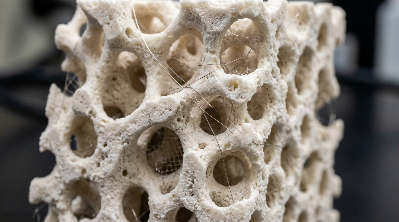
A scientific review in Bioelectronic Medicine reveals that electrical stimulation accelerates bone repair across complex clinical challenges, including delayed unions, non-unions, and critical-sized defects. Authored by Samira Farjaminejad and Aaron M. Dingle, the analysis synthesizes preclinical investigations and clinical trials showing how bioelectrical cues stimulate bone regeneration by modulating osteoblasts, osteoclasts, and mesenchymal stem cells. Although bone possesses an intrinsic capacity for self-repair, extensive structural voids and compromised fractures often fail to resolve naturally, requiring targeted orthopedic interventions. The review highlights how applied electrical fields enhance osteogenic differentiation, accelerate neovascularization, and drive extracellular matrix deposition, bridging cellular mechanotransduction with biochemical signaling cascades. By systematically evaluating bone’s biophysical properties alongside diverse stimulation modalities, the authors explain how electrical cues can overcome the persistent biological limitations of conventional bone autografts, allografts, and synthetic substitutes.
Natural bone functions as an electroactive composite material whose regeneration relies on coordinated cellular actions across three overlapping chronological phases: inflammation, repair, and remodeling. Farjaminejad and Dingle highlight that bone’s endogenous bioelectricity stems primarily from stress-induced piezoelectric phenomena first identified by Fukada and Yasuda. When physiological mechanical forces apply shear stress to bone’s extracellular framework, type I collagen fibrils undergo internal dipole realignment and charge displacement, generating localized electrical potentials across the tissue. Concurrently, the inorganic mineral phase composed of hydroxyapatite interacts directly with this collagen network, displaying semiconducting characteristics that support bioelectrical signal propagation and regulate ionic exchange, particularly the transport of calcium and phosphate ions vital for mineralization. These endogenous electrical fields serve as natural guidance cues that regulate the migration, proliferation, and differentiation of osteoblasts, osteoclasts, and mesenchymal stem cells, ensuring balanced structural renewal and skeletal load-bearing recovery.
At the cellular level, electrical stimulation operates by engaging membrane receptors and voltage-gated ion channels to translate physical electrical gradients into intracellular biochemical events. Voltage-gated calcium channels play a pivotal role by opening in response to membrane potential shifts, mediating calcium influx into osteogenic cells. In particular, L-type voltage-gated calcium channels direct calcium entry into mesenchymal stem cells and osteoblasts, activating osteogenic transcription factors such as Runx2 and Osterix while stimulating bone morphogenetic proteins and Wnt/beta-catenin pathways. Concurrently, P/Q-type and N-type calcium channels regulate osteoblast proliferation and govern calcium oscillatory rhythms in osteoclasts to control bone resorption. T-type calcium channels facilitate osteogenic differentiation and early osteoclastic fusion, whereas R-type channels support cytoskeletal remodeling, osteoblast adhesion, and osteoclast polarization. By elevating intracellular calcium levels and upregulating growth factors like vascular endothelial growth factor and transforming growth factor-beta, electrical stimulation accelerates both matrix mineralization and local angiogenesis.
To therapeutically harness these biophysical responses, clinicians utilize three primary electrical stimulation modalities, beginning with direct current electrical stimulation. Direct current stimulation involves implanting electrodes directly at the fracture or fusion site to deliver a continuous, low-intensity direct electrical current to surrounding bone tissue. Clinical investigations confirm that direct current stimulation provides substantial therapeutic value in promoting solid fusion during spinal fusion surgeries and resolving recalcitrant non-unions that resist conventional treatment.
As a non-invasive alternative, capacitive coupling electrical stimulation applies alternating electrical fields across injured skeletal regions through skin-placed external electrodes. By establishing an electric field near the fracture site, capacitive coupling modulates transmembrane potentials and stimulates intracellular calcium pathways, promoting osteogenic differentiation and enhancing osseointegration around orthopedic implants and spinal fusions. Computational modeling studies comparing sheep and human cadaver specimens showed that conventional external bone growth stimulators struggle to deliver therapeutic field intensities to deep fractures, an issue compounded by variable patient compliance during lengthy daily treatment regimens. To overcome tissue resistance without invasive open surgery, researchers have designed minimally invasive alternatives like the Injectrode, an injectable electrode system capable of delivering uniform electrical stimulation directly to deep fracture sites.
Pulsed electromagnetic field therapy represents an established non-invasive modality that employs external coils to induce localized electrical currents in bone through inductive coupling. Initially approved by the United States Food and Drug Administration in 1979 for treating non-unions, failed fusions, and congenital pseudoarthrosis, pulsed electromagnetic stimulation generates dynamic magnetic fields that stimulate osteoblast activity and promote vascularization without direct skin-to-electrode contact. Clinical trials indicate that pulsed electromagnetic fields shorten fracture healing times in long bones, osteotomies, and high-risk acute fractures. Nevertheless, variable clinical outcomes continue to appear across different studies due to disparities in magnetic waveforms, field strengths, treatment durations, and device calibration, emphasizing the need for standardized clinical protocols tailored to specific anatomical injury sites.
Published clinical trials provide quantitative evidence supporting pulsed electromagnetic field therapy across diverse orthopedic conditions. In femoral neck fractures managed with cancellous screws, adjunctive electromagnetic therapy produced a 94 percent successful healing rate compared to 69 percent in conventionally treated controls. In femoral component loosening following hip arthroplasty, electromagnetic stimulation achieved successful stabilization in 53 percent of cases versus only 11 percent among controls. For vertebral fractures, pulsed electromagnetic fields yielded a 92 percent successful healing rate compared to 65 percent in control cohorts. In avascular hip osteonecrosis, conservative electromagnetic treatment resulted in complete pain relief for 53 percent of patients and significant reduction for 26 percent, alongside healing rates of 81 percent in Steinberg stage II and 70 percent in stage III lesions. Moreover, electromagnetic therapy combined with mechanical stabilization achieved consolidation rates between 73 and 85 percent in established non-unions, demonstrating clear therapeutic efficacy.
Beyond standalone stimulation units, researchers are integrating electrical stimulation directly with advanced biomaterials and bone tissue engineering scaffolds. While traditional ceramic substitutes like hydroxyapatite, beta-tricalcium phosphate, and biphasic calcium phosphate provide osteoconductive architecture, they lack intrinsic bioelectrical activity. To overcome this limitation, bioengineers incorporate electroconductive polymers, including polypyrrole, polyaniline, and poly(3,4-ethylenedioxythiophene), to construct scaffolds that amplify electrical fields throughout the defect site. These conductive matrices accelerate mineral nucleation, enhance protein adsorption, improve calcium transport, and support mitochondrial activity. In vivo rodent experiments underscore the critical role of scaffold polarization: in Wistar rats, electrostatically poled polyvinylidene fluoride scaffolds generated significantly greater trabecular bone and bone marrow formation over four weeks than unpoled scaffolds, which produced no new bone. Combining electroactive scaffolds with biological factors, such as bone marrow mesenchymal stem cells and bone morphogenetic protein-2, further enhances osteogenic differentiation and vascular integration within large-volume defects.
Piezoelectric biomaterials represent an innovative category of electroactive scaffolds capable of generating electrical cues autonomously under physiological mechanical loading. Constructed from materials such as barium titanate nanoparticles, poly(L-lactic acid), lithium sodium potassium niobate, and collagen composites, these scaffolds convert mechanical stress into localized surface charges without external batteries or wiring. Laboratory investigations by Silva and colleagues revealed that dynamically stimulated piezoelectric scaffolds increase osteoblast proliferation by approximately 20 percent, driving substantial increases in matrix mineralization. By harnessing patient biomechanical motion to generate continuous electrical stimulation, self-powered piezoelectric scaffolds replicate bone’s native bioelectrical microenvironment, presenting an attractive therapeutic platform for critical-sized bone defects and complex reconstructive surgeries in orthopedic and maxillofacial medicine.
Despite these promising findings, significant translational hurdles impede the routine clinical deployment of electrical bone stimulation technologies. Preclinical rodent studies demonstrate positive osteogenic responses, but small-animal findings cannot readily predict human outcomes due to pronounced differences in bone thickness, tissue impedance, remodeling kinetics, and mechanical loading. Large-animal studies in sheep, pigs, and dogs provide greater clinical relevance, yet they confirm that non-invasive stimulators struggle to deliver adequate field strength across deep, dense skeletal structures. Regulatory frameworks also differentiate these technologies, with the Food and Drug Administration classifying non-invasive systems under product code LOF and implantable direct current stimulators under product code LOE. Overcoming these barriers will require interdisciplinary efforts integrating advanced computational modeling, machine learning to personalize stimulation parameters, injectable electrodes, and wireless bioelectronics. Standardizing treatment protocols and conducting rigorous longitudinal clinical trials will be essential to establish electrical stimulation as a mainstream therapeutic strategy in orthopedic regenerative medicine.
Subject of Research: Electrical stimulation mechanisms and therapeutic applications in bone regeneration
Article Title: The role of electrical stimulation in bone regeneration: mechanistic insights and therapeutic advances
Article References: Farjaminejad, S., & Dingle, A. M. (2025). The role of electrical stimulation in bone regeneration: mechanistic insights and therapeutic advances. Bioelectronic Medicine, 11(1), Article 18. https://doi.org/10.1186/s42234-025-00180-x
Image Credits: AI Generated
DOI: 10.1186/s42234-025-00180-x
Keywords: bone regeneration, electrical stimulation, piezoelectric scaffolds, bone tissue engineering, osteogenesis, PEMF therapy, bioelectronic medicine, orthopedic surgery, role, electrical, stimulation, bone
News Source: Denise Maddox. (November 8, 2026). How Electrical Stimulation and Bioelectrical Scaffolds Accelerate Bone Regeneration. Scienmag.