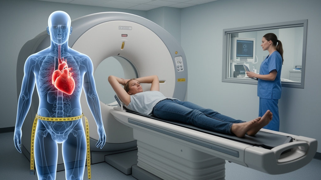
In a finding that could reshape how radiologists think about body composition and medical imaging, a new study has uncovered a surprising relationship between a person’s waist-to-hip ratio and the amount of radiation they receive during coronary CT angiography. The research, published in BMC Medical Imaging, suggests that individuals with higher waist-to-hip ratios — a measure of central obesity — actually receive lower radiation doses during these critical heart scans, primarily because the scans cover a shorter length of their body.
The study, led by Abdullah Şükün from the Department of Radiology at Başkent University Alanya Hospital in Turkey, along with colleagues from the hospital’s cardiology department, examined 134 consecutive adults who underwent coronary CT angiography. This non-invasive imaging technique uses computed tomography to visualize the coronary arteries and is a cornerstone in the diagnosis of coronary artery disease. The researchers wanted to understand whether simple anthropometric measurements — the kind taken routinely before these scans — could predict radiation exposure metrics reported by the scanner.
What makes this study particularly interesting is its focus on waist-to-hip ratio, or WHR, a metric that has received far less attention in imaging research than body mass index, or BMI. While BMI is a blunt instrument that only accounts for total body mass relative to height, WHR captures something more nuanced: the distribution of body fat, particularly the accumulation of visceral fat around the abdomen that is known to carry distinct metabolic and cardiovascular risks. The researchers hypothesized that this body shape measure might influence how scanners deliver and record radiation dose during cardiac imaging.
The team retrospectively analyzed data from 134 adults — 81 men and 53 women — with a mean age of 52.2 years. They extracted scanner dose reports and evaluated four key metrics: cardiac volumetric CT dose index (CTDIvol), cardiac-helical dose-length product (DLP), total examination DLP, and CCTA scan length. These metrics are standard measures of radiation exposure in CT imaging, with CTDIvol reflecting the radiation output per slice and DLP capturing the total radiation across the entire scanned volume.
The results revealed an unexpected pattern. Waist-to-hip ratio was inversely correlated with cardiac DLP — meaning higher WHR was associated with lower radiation dose-length product — and also with scan length, but notably not with CTDIvol. Specifically, each 0.1-unit increase in WHR was associated with a 28.3 mGy·cm reduction in cardiac DLP and a 0.67 cm shorter scan length. These associations held up even after adjusting for body surface area, age, sex, pre-scan heart rate, and beta-blocker administration.
To understand why this counterintuitive relationship exists, the researchers turned to a sophisticated statistical technique called path-based decomposition. This method allowed them to mathematically separate the direct effect of WHR on radiation dose from the indirect effect that operates through scan length. The results were striking: nearly 89 percent of the total association between WHR and cardiac DLP was explained by the indirect pathway through scan length. In other words, people with higher waist-to-hip ratios received shorter scans, and shorter scans naturally meant lower total dose-length products.
This finding has important implications for how we think about radiation dosimetry in cardiac imaging. The dose-length product, by its very mathematical definition, incorporates both the intensity of radiation and the longitudinal extent of tissue being irradiated. So when scan length shrinks, DLP inevitably falls — regardless of whether the radiation intensity per unit length changes at all. The study’s authors were careful to emphasize this point, noting that their findings describe a statistical relationship rather than an independently established causal mechanism.
Indeed, the researchers explicitly caution against overinterpreting their results. They state that these findings do not justify WHR-guided protocol changes without prospective validation. This is a crucial distinction in medical research — observing that two variables are statistically associated, even through a well-characterized indirect pathway, is fundamentally different from establishing that one variable causally drives the other in a way that should inform clinical practice.
The study also uncovered an independent association between pre-scan heart rate and CTDIvol, with each beat-per-minute increase associated with a 0.25 mGy decrease in CTDIvol. This finding likely reflects the clinical reality that patients with higher heart rates may be scanned using different protocols or with different tube current settings, though the exact mechanism remains speculative without further investigation.
Mean radiation dose metrics in the study population were 45.7 mGy for cardiac CTDIvol, 708.2 mGy·cm for cardiac DLP, 789.9 mGy·cm for total examination DLP, and a mean scan length of 12.0 cm. These values provide a useful reference point for understanding the magnitude of radiation exposure in contemporary coronary CT angiography practice and how anthropometric variables might modulate them.
As medical imaging continues to evolve toward more personalized protocols, studies like this one highlight the complex interplay between patient anatomy, scanner technology, and radiation exposure. While the waist-to-hip ratio may not yet be ready for prime-time clinical decision-making in radiology suites, the research underscores the value of looking beyond BMI to more nuanced measures of body composition. The path forward will require prospective studies that can establish causal relationships and determine whether anthropometric measurements can genuinely inform protocol optimization — a goal that remains tantalizingly within reach as imaging technology and statistical methodologies continue to advance together.
Subject of Research: The relationship between waist-to-hip ratio and radiation dose metrics in coronary CT angiography
Article Title: Association between waist-to-hip ratio and radiation dose metrics in coronary CT angiography
Article References: Şükün, A., Doğmuş, F., Akbay, E., Akıncı, S., & Adar, A. (2026). Association between waist-to-hip ratio and radiation dose metrics in coronary CT angiography. BMC Medical Imaging. https://doi.org/10.1186/s12880-026-02770-w
Image Credits: AI Generated
DOI: 10.1186/s12880-026-02770-w
Keywords: coronary CT angiography, radiation dose, waist-to-hip ratio, anthropometry, dose-length product, scan length, body composition, medical imaging, CTDIvol, cardiovascular imaging, radiology, obesity
News Source: Ophelia Keating. (October 18, 2026). How Body Shape Shapes Your Radiation Dose: Waist-to-Hip Ratio Emerges as Unexpected Factor in Heart CT Scans. Scienmag.