


Scientists at the University of Miami have mapped out why a small pad of fat hidden deep inside the hip joint could become the next frontier in regenerative medicine for osteoarthritis, while also cautioning that the evidence is still too thin to call it a therapy. In a narrative review published in Frontiers in Bioengineering and Biotechnology, the team compared the cotyloid fossa fat pad of the hip with its far better-studied counterpart in the knee, the infrapatellar fat pad, and found a striking imbalance: one tissue has been dissected down to its stem cells and secreted molecules, while the other remains largely a biological blank spot despite sitting directly beside the cartilage it may influence.
Hip osteoarthritis is a leading cause of pain and disability worldwide, yet no validated disease-modifying orthobiologic therapy currently exists for the hip joint. That gap motivated the review, led by R. P. Rossow and Dimitrios Kouroupis of the Department of Orthopaedics at UHealth Sports Medicine Institute and the Diabetes Research Institute at the Miller School of Medicine. Their central argument is that the knee’s infrapatellar fat pad, or IFP, offers a rigorous reference model against which the hip’s cotyloid fossa fat pad, the CFFP, can now be systematically tested, rather than assumed to behave the same way.
The IFP, also known as Hoffa’s fat pad, sits behind the patellar tendon in the knee and has been recharacterized over the past decade from a passive cushion into an active immunological and metabolic organ. It contains macrophages, fibroblasts, adipocytes, endothelial cells and resident mesenchymal stem or stromal cells, known as MSC. During osteoarthritis, IFP macrophages shift toward a pro-inflammatory M1-like state and release cytokines, prostaglandins and adipokines such as IL-6, TNF-alpha and leptin. These mediators correlate with cartilage thinning and patient-reported symptoms, and as inflammation persists the pad becomes fibrotic and more vascularized, stiffening the tissue and increasing mechanical stress within the joint.
What makes the IFP especially valuable as a model is the depth of characterization its MSC population has undergone. Flow cytometry studies report that more than 95 percent of IFP-MSC express the canonical identity markers CD73, CD90 and CD105, while CD34, CD45, CD56 and HLA-DR are nearly absent. The cells also carry high levels of CD10, an enzyme also called neprilysin that degrades Substance P, a neuropeptide that drives inflammatory and nociceptive signaling. In preclinical models, this Substance P degradation capacity has been shown to reverse synovitis and fat pad fibrosis. IFP-MSC additionally modulate Calcitonin Gene-Related Peptide-associated neurogenic inflammation, and their secretome, spanning immunomodulatory cytokines, growth factors and microRNAs carried in extracellular vesicles, has been profiled in detail.
The therapeutic toolkit built around IFP-MSC extends further. Priming strategies, including hypoxic culture, cytokine-based inflammatory priming with interferon-gamma and TNF-alpha, and hormonal priming with oxytocin, have been shown to enhance immunomodulatory potency in vitro, pushing the cells toward an immunosuppressive phenotype marked by IDO, HLA-G, PD-L1, prostaglandin E2 and TSG-6. Engineered versions of the cells, transduced with a CGRP antagonist gene, produce extracellular vesicles with enhanced analgesic and anti-inflammatory cargo aimed at neurogenic pain pathways. Early human studies of intra-articular IFP-MSC products in knee osteoarthritis report safety and preliminary improvements in pain and function, though the review stresses that large-scale randomized controlled trials are still lacking.
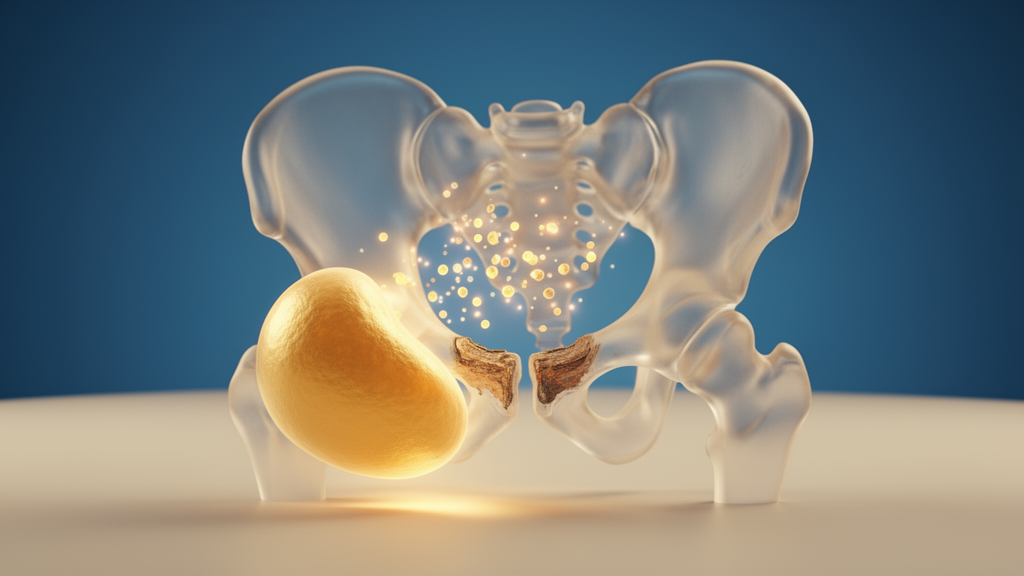
Against this mature evidence base, the CFFP column of the comparison is strikingly sparse. The cotyloid fossa is a hollow at the center of the acetabulum, composed of adipose and synovial tissue that envelops the ligamentum teres and contributes to hip stability, lubrication and load distribution. Once regarded as a passive space-filling structure, it is now recognized as an active participant in joint physiology, housing a vascularized stromal compartment with resident immune cells and mesenchymal progenitors. Studies have identified MSC-like populations there with self-renewal and osteogenic and chondrogenic differentiation potential, and recent work characterizing human pulvinar sub-synovial adipose tissue across aging and osteoarthritis reported a disease-associated expansion of DPP4-positive and THY1-positive mesenchymal progenitor cells.
Yet the review is explicit about what has not been done. No flow cytometry panel, no colony-forming assay and no secretome profiling has been reported from directly isolated CFFP tissue. No study has compared CFFP-MSC with IFP-MSC under inflammatory conditions, tested whether hip-resident cells support CD10 enrichment and neuropeptide modulation, or characterized the priming responses or extracellular vesicle function of the hip population. Inflammatory mediator profiling of the CFFP remains limited, and much of the existing evidence comes from studies of pulvinar synovium or intra-articular adipose tissue rather than the fat pad itself. The authors conclude that CFFP-MSC and CFFP-derived extracellular vesicles cannot yet be presented as validated hip-specific orthobiologic candidates.
The clinical correlates that do exist are observational. In hip osteoarthritis, inflammation of the cotyloid fossa synovium correlates with labral pathology and functional disability, and cotyloid fossa dimensions correlate with labral tear size and SF-12 scores. However, this does not appear to scale directly with fat pad size or gross morphology, suggesting that inflammatory signaling at the site may arise from mechanical insult or secondary synovial activation rather than constitutive metabolic activity. The authors argue this distinction has fueled a view of the CFFP as less biologically active than the IFP, a conclusion that may reflect an evidence gap rather than a true biological difference. Kuhns and colleagues documented inflammatory and remodeling changes in pulvinar synovial tissue through biochemical and histologic analysis of patients with femoroacetabular impingement, supporting the plausibility of fat-synovium-immune interactions in the hip.
The review also injects a note of caution drawn from the knee experience. Extracellular vesicle effects can be heterogeneous: a recent study showed that a mitochondria-derived vesicle subpopulation from IFP-MSC transferred mitochondrial DNA to chondrocytes, activated cGAS-STING signaling and worsened cartilage degradation and synovitis in a rat osteoarthritis model, with STING inhibition reversing the damage. Donor disease state, vesicle subtype and cargo composition influenced how harmful the preparation was, and the pathogenic subpopulation was enriched in osteoarthritic joints. The authors argue that any future CFFP vesicle therapy will require subpopulation-level profiling and fractionation rather than bulk isolation, since a whole-population preparation could carry harmful cargo that bulk characterization would miss.
The translational roadmap the authors propose begins with baseline characterization of CFFP-MSC through surface phenotyping, clonogenic capacity, multilineage potential and basal immunomodulatory gene expression, followed by head-to-head functional comparison with IFP-MSC under controlled inflammatory stimuli, applying the cytokine, oxytocin and hypoxia priming paradigms already established for knee cells. Donor factors such as age, sex, obesity and disease stage, which are known to shape fat pad MSC biology, should be matched or reported to avoid confounding tissue-of-origin differences. If hip-resident cells do carry the immunomodulatory signatures of their knee counterparts, the authors suggest they would represent an endogenous, arthroscopically accessible and disease-relevant source for early joint-preserving treatment, but they emphasize that this remains a testable hypothesis, that the comparative biology must precede therapeutic development, and that no current evidence establishes superiority of hip-resident cells over other MSC sources.
Subject of Research: Comparing the hip's cotyloid fossa fat pad with the knee's infrapatellar fat pad as sources of mesenchymal stem cells and extracellular vesicles for osteoarthritis therapy
Article Title: The cotyloid fossa fat pad as a hip-specific orthobiologic niche: lessons from the infrapatellar fat pad for mesenchymal stem/stromal cell and extracellular vesicle therapy in osteoarthritis
Article References: Rossow, R. P., Polavarapu, K., Naik, M., Kaplan, L. D., Best, T. M., Rizzo, M. G., & Kouroupis, D. (2026). The cotyloid fossa fat pad as a hip-specific orthobiologic niche: lessons from the infrapatellar fat pad for mesenchymal stem/stromal cell and extracellular vesicle therapy in osteoarthritis. Frontiers in Bioengineering and Biotechnology, 14, Article 1930127. https://doi.org/10.3389/fbioe.2026.1930127
Image Credits: AI Generated
DOI: 10.3389/fbioe.2026.1930127
Keywords: osteoarthritis, mesenchymal stem cells, extracellular vesicles, infrapatellar fat pad, cotyloid fossa fat pad, hip joint, synovium, immunomodulation, orthobiologics, cartilage, regenerative medicine, tissue engineering
News Source: Drew Townsend. (October 28, 2026). Hip fat pad may hold key to stem cell therapy for osteoarthritis. Scienmag.



