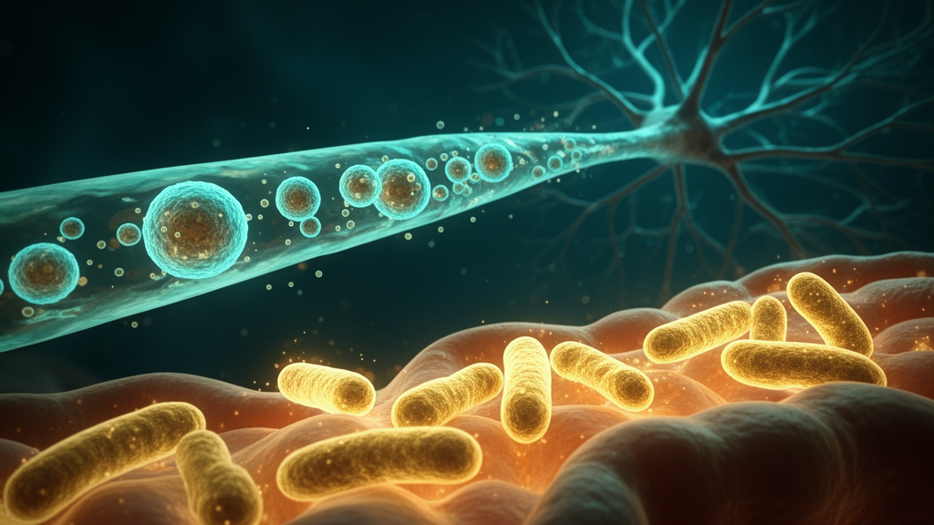
A mini review published in Frontiers in Microbiology synthesizes preclinical evidence demonstrating that the gastrointestinal tract functions as a primary educational hub for the systemic immune system. Carolina Prado and Daniel Hevia-Magaña argue that the classical paradigm of central nervous system immune privilege is evolving into a dynamic, multi-organ neuroimmune axis. In this model, the gastrointestinal mucosa acts not merely as a localized physical barrier, but as a vital center that instructs systemic immune responses. Dietary inputs, microbial metabolites, and commensal colonization imprint intestinal adaptive and innate immune cells with distinct functional phenotypes and migratory signatures, preparing them for deployment to distant sites, including the brain.
The review highlights that under homeostatic conditions, gut-derived signals direct the physiological seeding of central border zones and brain parenchyma. Specialized immune populations maintain baseline tissue surveillance, reinforce barrier integrity, and calibrate neurodevelopmental behaviors. Conversely, the authors note that pathobiont expansion, the loss of short-chain fatty acid-producing commensals, and mucosal barrier disruption predominantly promote pathogenic gut priming. These perturbations transform gut-resident cells into autoreactive or pro-inflammatory effectors that breach central neurovascular borders, contributing to neurodegeneration, ischemic injury, and neuroinflammatory pathology.
Early-life microbial colonization is critical for this process, as the thymus participates in active enterothymic communication. In murine models, colonization during the neonatal window enables colon-derived immune populations, primarily plasmacytoid dendritic cells and CD3+ T-cells, to migrate to the thymus. This baseline distribution process is disrupted if early-life dysbiosis is induced via antibiotic exposure, with effects persisting into adulthood. Intestinal colonization promotes the physical trafficking of microbial antigens from the intestine directly to the thymus by migrating intestinal dendritic cells, which coordinates the expansion and selection of a microbiota-specific T cell repertoire.
Recent animal studies demonstrate that the maturation of the gut microbiota and the establishment of steady-state brain T cells are functionally synchronized during weaning. Upon the dietary transition to solid food, the resulting shift toward a mature microbiome is associated with the egress of gut-educated CD4+ T cells from the gastrointestinal tract into the bloodstream. In murine models, these cells subsequently migrate to brain parenchyma compartments, such as the subfornical organ, where they contribute to regulating homeostatic behavior via IFNγ signaling. Germ-free models completely lack this parenchymal T cell niche, illustrating that the physiological colonization of the CNS by adaptive immune cells is directly dependent on chronological gut-microbiome maturation during weaning.
Metabolic imprinting by short-chain fatty acids (SCFAs) further shapes this immune repertoire. SCFAs are synthesized via anaerobic fermentation of non-digestible complex carbohydrates and dietary fibers. Within the gut-associated lymphoid tissue, these metabolites imprint intestinal lymphoid and myeloid cells with specific homeostatic and tolerogenic programming. SCFA binds to specific G protein-coupled receptors, including FFAR2, FFAR3, and GPR109A, which are abundantly expressed on intestinal epithelial cells, dendritic cells, and regulatory T cells. Activation of these receptors on colonic dendritic cells and macrophages induces protective anti-inflammatory pathways while suppressing pro-inflammatory transcriptional hubs such as the NF-κB pathway.
Additionally, butyrate and propionate act as natural class I and II histone deacetylase inhibitors after being transported into immune cells via monocarboxylate transporters. This intracellular inhibition significantly augments histone H3 acetylation at the promoter and conserved non-coding sequence 1 regions of the Foxp3 locus, which is essential for the stabilization and generation of regulatory T cells. The functional consequence of this synchronized signaling is a profound induction of regulatory populations within the gut-associated lymphoid tissue, destined for peripheral deployment. SCFAs directly promote the differentiation, survival, and clonal expansion of mucosal FoxP3+ regulatory T cells, enhancing their suppressive capacity and priming them for systemic tissue tropism.
Pathogenic priming represents a distinct cellular program that must be conceptually separated from simple leaky gut or passive bacterial translocation. Gut priming involves an active, highly orchestrated shift in the phenotypic identity and homing profile of mucosal immune cells. Within the adaptive compartment, colonization by Segmented Filamentous Bacteria drives local differentiation of Th17 cells. These gut-primed Th17 cells acquire a stem-like memory phenotype, allowing them to circulate systemically and eventually differentiate into pathogenic effectors in distal tissues. During systemic transit, these cells exploit molecular mimicry, a process where T cells initially trained against commensal microbial antigens cross-react with structurally similar self-antigens embedded within extra-intestinal target tissues.
In the context of the gut-brain axis, this is a proposed contributor to CNS autoimmunity. Although retinoic acid-dependent gut education primarily upregulates the α4β7 integrin, it also drives expression of the shared α4 subunit, allowing pleiotropic pairing with β1 to form α4β1. Because its primary ligand, vascular cell adhesion molecule 1, is heavily expressed on central neurovascular boundaries and the blood-brain barrier during inflammation, this aberrant dual homing profile allows gut-educated, myelin-reactive Th17 cells to actively patrol, infiltrate, and disrupt the CNS parenchyma. This mechanism is observed in experimental autoimmune encephalomyelitis models and is inferred in human multiple sclerosis.
Pathogenic priming is not exclusive to adaptive lymphocytes; physical disruptions to the mucosal barrier and localized tissue stress directly instruct innate populations for remote recruitment. Severe intestinal barrier breakdown and subsequent inflammatory cues activate gut-resident macrophages via Triggering Receptor Expressed on Myeloid cells 1 signaling. This acute stimulus actively rewrites the functional programming of these phagocytes, prompting their phenotypic conversion and enabling their egress from the lamina propria. Once mobilized, these gut-derived myeloid cells cross the compromised blood-brain barrier and infiltrate the brain parenchyma, where they amplify ischemic injury and perpetuate neuroinflammation.
The transition of immune cells from the confined environment of the intestinal mucosa to systemic sites is a highly regulated process involving specific sphingolipid gradients, chemokine receptors, and anatomical routes. The Sphingosine-1-phosphate gradient serves as a critical regulator of lymphocyte egress from intestinal tissues. Most gut-educated cells, including B-cells, T cells, and innate lymphoid cells, rely on the expression of the S1P receptor to sense the higher concentrations of S1P in the lymph node and blood compared to the tissue. High-resolution photolabeling has confirmed that this pathway acts as a key mechanical license for adaptive lymphocytes to exit mucosal boundaries. However, myeloid-cell trafficking is more context- and subset-dependent, integrating chemokine-receptor signaling, tissue-derived inflammatory cues, and locally generated pro-resolving mediators rather than relying on a single universal pathway.
Once leukocytes leave the gut, they redistribute through distinct anatomical routes dictated by their cellular identity, activation status, and microenvironmental cues. Traditional immunological dogmas dictate that tissue-emigrating cells enter afferent lymphatic vessels to populate regional draining lymph nodes, such as the mesenteric lymph nodes. Cell tracking studies establish that this classic route is highly favored by antigen-presenting cells and specific memory T cell subsets executing classical immune surveillance loops. Intriguingly, high-resolution endoscopic photoconversion tracking has revealed that a substantial proportion of gut-emigrating immune cells, heavily dominated by B cells and unconventional or innate-like T cell populations, subsequently seeds systemic reservoirs like the spleen. However, the detection of gut-photoconverted immune cells in the spleen demonstrates the systemic redistribution of cells previously located in the labeled intestinal compartment, rather than defining a direct vascular conduit.
To demonstrate that gut immune cells physically relocate between these anatomically distant systems, modern neuroimmunology relies heavily on photoconvertible reporter mouse models. These models express engineered fluorescent proteins that, upon brief exposure to violet or ultraviolet light, irreversibly shift from green to red emission. In gut-brain axis research, targeted surgical or endoscopic intraluminal illumination photo-tags intestinal populations locally within the gut wall. This tissue-specific labeling enables researchers to trace the relocation of gut-educated immune cells as they leave the lamina propria, navigate systemic vascular networks, and populate distinct CNS compartments under homeostatic or pathological conditions. By confirming prior tissue origin rather than continuously resolving every intermediate dynamic mechanism in vivo, photoconversion establishes a spatial link between intestinal education and central infiltration.
While direct cell-tracking in human CNS tissue remains methodologically restricted, human post-mortem studies and cerebrospinal fluid profiling complement these rodent paradigms by confirming the presence of gut-reactive lymphocytes at neurovascular boundaries. Clinical analyses of cerebrospinal fluid and brain tissue samples from multiple sclerosis patients during active neuroinflammatory crises confirm the presence and protective anti-inflammatory role of gut-derived IgA+ plasma cells. These cells locally secrete IL-10 and IgA to suppress encephalitogenic T-cell expansion and prevent brain parenchyma breach. The review concludes that translating these cellular dynamics into clinical application requires addressing significant translational and methodological challenges, including the heavy reliance on animal models and the inability to perform real-time cell labeling in humans. Key species differences in gut microbiota composition, intestinal microarchitecture, and blood-brain barrier dynamics make it difficult to determine the exact quantitative contribution of gut-derived leukocytes to human neuroinflammatory pathology.
Subject of Research: Microbiota-driven immune trafficking along the gut-brain axis
Article Title: From gut education to central infiltration: microbiota-driven immune trafficking along the gut-brain axis
Article References: Prado, C., & Hevia-Magaña, D. (2026). From gut education to central infiltration: microbiota-driven immune trafficking along the gut-brain axis. Frontiers in Microbiology, 17, Article 1986940. https://doi.org/10.3389/fmicb.2026.1986940
Image Credits: AI Generated
DOI: 10.3389/fmicb.2026.1986940
Keywords: gut-brain axis, neuroimmune axis, immune trafficking, gut-associated lymphoid tissue, short-chain fatty acids, regulatory T cells, Th17 cells, macrophages, S1PR1 signaling, photoconversion tracking, meningeal immunity, blood-brain barrier, neuroinflammation, microbial metabolites, intestinal barrier, TREM1 signaling, molecular mimicry, homeostatic seeding, pathobiont expansion, CNS immune privilege
News Source: Cassandra Pierce. (November 14, 2026). Gut microbes shape immune cells that migrate to the brain. Scienmag.