The trillions of microbes living in the human gut change dramatically as we age, and a growing body of research suggests those changes are not merely a byproduct of getting old but active participants in the decline of intestinal health. A new study published in Aging Cell has taken one of the most direct approaches yet to testing that idea: researchers transplanted fecal microbiota from healthy elderly people and from young adults into young pigs, then tracked what happened to the animals’ gut structure, barrier function, blood chemistry, and gene expression. The results show that the age of the donor leaves a measurable imprint on the recipient’s intestine, with microbiota from elderly donors weakening the gut lining, shifting the plasma metabolome, and suppressing a coordinated module of antiviral defense genes in the intestinal mucosa.
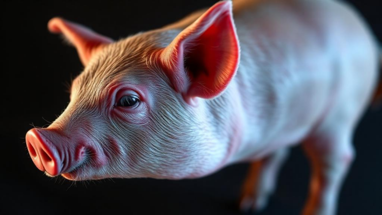
The choice of the pig as the experimental recipient was central to the study’s design. Pigs share striking anatomical and molecular similarities with humans, including comparable ratios of small intestine length to body weight, well-developed submucosal glands and villus structures, similar cell populations, and a high degree of genomic homology. Previous comparative work has also shown that mature human donor microbiota establish more efficiently and persistently in piglets than in mice under matched conditions. That makes the porcine intestine a valuable translational bridge between reductionist rodent experiments and human intestinal biology, particularly for a question that hinges on how an intact human microbial community behaves in a mammalian gut.
To carry out the experiment, the team worked with eighteen two-month-old Bama miniature pigs, divided into a control group and two groups receiving fecal microbiota from either young donors aged 22 to 29 years or elderly donors aged 70 to 85 years. Five donors contributed to each age group, with two fecal samples per donor. The researchers first depleted the pigs’ resident microbes with a three-day course of oral antibiotics, then administered human fecal suspensions on alternate days over eight days. After a week of colonization, they collected blood, intestinal tissues, and gut contents for analysis. The antibiotic pretreatment sharply reduced microbial diversity, and transplantation restored overall richness to baseline levels, but the compositional structure of the community was substantially reshaped by the transplanted human microbiota, with clear separation between FMT recipients and control animals.
Microbial profiling revealed how profoundly donor age shaped the reconstituted communities. Although pigs receiving young and elderly donor microbiota showed no significant differences in alpha diversity, meaning overall richness and evenness, their community structures differed significantly. Pigs colonized with elderly donor microbiota exhibited a reduced gut microbiome health index and an elevated microbial dysbiosis index, exploratory measures originally derived from human datasets. The two age groups also produced distinct core communities: 986 amplicon sequence variants were unique to the young donor group and 797 to the elderly donor group, with only 611 shared. Notably, pigs receiving young donor microbiota harbored higher proportions of transient microbial variants, suggesting greater environmental sensitivity, while communities from elderly donors contained fewer such variants, implying reduced responsiveness to environmental fluctuations. At the species level, Phocea massiliensis was enriched in pigs receiving young donor microbiota, whereas Blautia obeum emerged as a signature species in those receiving elderly donor microbiota.
These microbial differences translated into structural and functional consequences for the gut. Histological examination showed that ileal villus height was significantly reduced in pigs transplanted with elderly donor microbiota, while the duodenum, jejunum, and colon were largely spared, pointing to a region-specific impact of donor age on intestinal architecture. Reduced villus height is significant because it may partly account for the impaired nutrient absorption frequently reported in older adults, and the accompanying transcriptomic data reinforced this connection, with differential gene expression enriched in pathways related to lipid digestion and absorption. More striking still were the barrier findings: serum diamine oxidase, a marker of mucosal disruption and intestinal permeability, was significantly elevated in pigs receiving elderly donor microbiota, and levels of the tight junction proteins ZO-1, Claudin-1, and Occludin were markedly reduced across the jejunum, ileum, and colon.
Correlation analysis linked specific bacterial species to these barrier changes. The abundance of P. massiliensis was positively correlated with ileal Claudin-1 expression, while B. obeum abundance was negatively associated with tight junction protein expression across multiple intestinal segments. This pattern is intriguing given what is known about the two organisms. P. massiliensis, a strict anaerobic Gram-negative bacillus found in the intestines of humans and animals, has shown age-associated decline in mouse models, and murine studies have reported a negative correlation between frailty and its abundance. B. obeum, by contrast, has been implicated as a potential risk factor in several diseases, including the exacerbation of colitis in mice, and its increased abundance has been associated with altered markers of diabetes risk. The enrichment of B. obeum in the elderly gut may therefore predispose individuals to age-related disease susceptibility, although the authors caution that these relationships remain correlative.
The metabolomic analysis added another layer to the story. Plasma metabolic signatures separated clearly between the two FMT groups, and five metabolites differed significantly: 3-methyloxindole, prostaglandin E3, and 2-hydroxybutanoic acid were elevated in pigs receiving elderly donor microbiota, while 2-hydroxyoctadecanoic acid and the dipeptide Tyr-Phe were diminished. The three elevated metabolites were inversely correlated with tight junction protein expression across intestinal segments, whereas the two reduced metabolites showed positive correlations with selected barrier proteins. To probe function directly, the researchers tested these metabolites on porcine intestinal epithelial cells in vitro. Tyr-Phe increased both cell viability and transepithelial electrical resistance, a measure of barrier tightness, providing evidence for a protective role. 2-Hydroxyoctadecanoic acid promoted cell viability but did not alter resistance under the tested conditions. Prostaglandin E3, meanwhile, left viability unchanged but reduced transepithelial electrical resistance, consistent with earlier work showing that prostaglandin E3 increases paracellular permeability in human intestinal cell monolayers through EP1- and EP4-mediated signaling.
Transcriptomic profiling of the ileal mucosa revealed 108 genes significantly upregulated and 109 downregulated in pigs receiving elderly donor microbiota, with enrichment in pathways including steroid hormone biosynthesis, retinol metabolism, PPAR signaling, and bile secretion. Each of these pathways has plausible relevance to intestinal aging: retinoid metabolism is closely tied to epithelial homeostasis and mucosal immune regulation, PPAR signaling governs energy metabolism and oxidative stress responses, and bile acids are microbiota-modified signaling molecules that regulate epithelial renewal and permeability. The most striking finding, however, emerged from hub gene analysis. The top seven genes identified by network analysis, MX2, ISG15, IFI6, IFIT1, OAS1, DHX58, and ISG12(A), were all significantly downregulated in pigs receiving elderly donor microbiota, and all belong to a coherent module of interferon-stimulated genes that form the intestine’s baseline antiviral defense.
This coordinated suppression of the interferon-stimulated gene module carries particular weight because tonic interferon signaling in the gut is not merely a response to infection but contributes to basal mucosal preparedness. Commensal bacteria can stimulate localized interferon-lambda-dependent expression of these genes in intestinal epithelial cells, establishing preemptive antiviral defense. The downregulated genes span multiple levels of the antiviral cascade: MX2 blocks viral replication after entry, IFIT1 recognizes non-self viral RNA, OAS1 activates an RNA-degrading antiviral pathway, and DHX58 modulates viral RNA sensing. Several of these genes, including ISG15, IFI6, and ISG12(A), also positively correlated with ileal tight junction protein expression, suggesting a mechanistic link between the suppression of epithelial interferon responsiveness and impaired barrier integrity. Elderly donor microbiota may either provide weaker microbial cues for maintaining this homeostatic antiviral tone or generate signals that dampen epithelial interferon responsiveness.
The authors are careful to frame their findings as association-based rather than definitively causal, noting that the study’s comparative design, moderate sample size, and short observation period leave open questions about the specific contributions of individual taxa, metabolites, and host genes. Future work, they suggest, should test whether aged microbiota directly suppress epithelial interferon signaling, whether supplementation with young donor microbiota or defined metabolites restores antiviral gene expression, and whether manipulating the interferon axis can rescue barrier function. Even with those caveats, the study delivers a compelling demonstration that the chronological age of a gut microbial community can reprogram the biology of its host, from villus architecture to plasma metabolites to mucosal gene expression. As populations worldwide age, the prospect of microbiota-targeted nutritional or therapeutic strategies designed to preserve intestinal integrity in the elderly looks increasingly grounded in mechanism rather than mere correlation.
Subject of Research: Effects of age-specific human gut microbiota transplantation on intestinal barrier function, metabolism, and mucosal gene expression in pigs
Article Title: Transplantation of Elderly Human Gut Microbiota Into Pigs Reprograms Intestinal Barrier Function, Plasma Metabolome, and Gut Mucosal Transcriptomic Landscape
Article References: Wang, J., Li, L., Mei, L., Tang, Z., Liao, B., Zhao, Q., Wang, Y., Fu, Q., Ren, L., Zhai, Z., Xu, Y., Yang, A., Duan, S., Zhai, Z., Hao, Y., Zhou, Y., Xu, Y., Yang, Y., Wu, Z., & Ji, Y. (2026). Transplantation of Elderly Human Gut Microbiota Into Pigs Reprograms Intestinal Barrier Function, Plasma Metabolome, and Gut Mucosal Transcriptomic Landscape. Aging Cell, 25(10), Article e70725. https://doi.org/10.1111/acel.70725
Image Credits: AI Generated
DOI: 10.1111/acel.70725
Keywords: gut microbiota, fecal microbiota transplantation, aging, intestinal barrier, tight junctions, pigs, metabolomics, transcriptomics, interferon-stimulated genes, Blautia obeum, Phocea massiliensis, intestinal permeability
Cite Scienmag News
APA
MLA
Chicago
Morgan Morrow. (October 2, 2026). Gut Microbes From Elderly Humans Weaken Intestinal Barriers When Transplanted Into Pigs. Scienmag. https://scienmag.com/gut-microbes-from-elderly-humans-weaken-intestinal-barriers-when-transplanted-into-pigs/
Morgan Morrow. “Gut Microbes From Elderly Humans Weaken Intestinal Barriers When Transplanted Into Pigs.” Scienmag, 2 October 2026, https://scienmag.com/gut-microbes-from-elderly-humans-weaken-intestinal-barriers-when-transplanted-into-pigs/. Accessed 2 October 2026.
Morgan Morrow. “Gut Microbes From Elderly Humans Weaken Intestinal Barriers When Transplanted Into Pigs.” Scienmag. October 2, 2026. https://scienmag.com/gut-microbes-from-elderly-humans-weaken-intestinal-barriers-when-transplanted-into-pigs/
Copy citation
Download RIS
Tags: age-related changes in gut microbiotaAgingBlautia obeumcross-species microbiota transfereffects of elderly microbiota on gut structureelderly human fecal microbiota transplantationfecal microbiota transplantationgut healthgut microbiome and aginggut microbiotainterferon-stimulated genesintestinal barrierintestinal barrier function declineintestinal permeabilityMetabolomicsmicrobiome immunologymicrobiome influence on gut healthmicrobiome-driven metabolic shiftsmicrobiota and antiviral gene suppressionmicrobiota transplantation and intestinal integrityPhocea massiliensispig models for human microbiome studiespigstight junctionsTranscriptomics