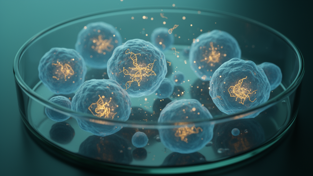
Imagine a molecule so small that it cannot be seen under a conventional microscope, yet so programmable that it can switch on a fluorescent beacon inside a living cancer cell the moment it finds its target. That is the promise of fluorogenic RNA aptamers, short engineered RNA strands that activate the fluorescence of otherwise dark small-molecule dyes upon binding. A new open-access mini-review published in Molecular Biology Reports by Mohammad Mehdi Akbarin, Zahra Farjami, and Hugo Ramírez Álvarez of the National Autonomous University of Mexico surveys this rapidly expanding field and argues that these glowing RNA molecules are emerging as central platforms for molecular imaging, biosensing, and precision oncology. The review, which synthesizes structural biology, fluorophore chemistry, and biosensor engineering across brain, breast, colorectal, liver, lung, prostate, and skin cancers, arrives at a moment when the technology is transitioning from elegant proof-of-concept experiments toward clinically relevant diagnostic tools.
The core principle behind fluorogenic RNA aptamers is deceptively simple. In solution, fluorogenic dyes such as DFHBI, the ligand recognized by the Spinach aptamer, are nearly non-fluorescent because they undergo rapid intramolecular rotation that dissipates absorbed energy as heat rather than light. When an aptamer folds into its defined three-dimensional structure, it creates an organized binding pocket that physically constrains the dye, restricting its conformational motion and stabilizing a planar fluorescent state through π–π stacking and hydrogen-bonding interactions. This reduces non-radiative decay and dramatically increases the fluorescence quantum yield. Because the RNA itself is genetically encodable, cells can be programmed to produce these light-up probes at specific genomic loci, enabling real-time visualization of RNA molecules, gene expression, and dynamic cellular processes in living systems without the need for fluorescent proteins.
The field originated with Spinach, the first widely adopted fluorogenic RNA aptamer, which binds DFHBI and produces green fluorescence analogous to green fluorescent protein. Although Spinach demonstrated the feasibility of genetically encoded RNA imaging, it suffered from folding instability, thermal sensitivity, and reduced fluorescence under physiological conditions. Successive generations were engineered to overcome these limitations: Spinach2 and Baby Spinach improved folding efficiency, the Broccoli family enhanced intracellular stability, and structurally distinct families including Mango, Corn, Pepper, Squash, and Beetroot each introduced unique fluorogen specificities, spectral properties, and structural architectures. Mango aptamers, for instance, exhibit high fluorescence brightness and strong ligand affinity through G-quadruplex-mediated fluorogen stabilization, while Corn produces yellow fluorescence with improved photostability, Pepper enables multicolor imaging through HBC fluorogens, and Beetroot expands the spectral range with red-shifted fluorescence, facilitating multiplex imaging in combination with other aptamers.
Structural analyses have revealed that fluorogenic RNA aptamers share common architectural principles despite their sequence diversity. Following transcription, these RNAs fold into complex secondary and tertiary conformations involving stem-loops, junctions, non-canonical base pairs, pseudoknots, and long-range interactions that generate precisely organized ligand-binding pockets. Several families, including members of the Mango and Spinach lineages, contain G-quadruplex motifs that provide rigid structural scaffolds for fluorogen binding. However, G-quadruplex formation is not a universal requirement: Pepper adopts a non-G-quadruplex architecture for HBC recognition, whereas Beetroot uses a compact RNA fold to bind DFAME. Crystallographic studies of Spinach, Spinach2, Mango, Corn, and Squash have provided direct evidence for how these pockets position fluorogens within constrained environments, while NMR spectroscopy and molecular dynamics simulations have revealed that aptamers can populate multiple conformational states during folding and ligand binding, explaining why fluorescence intensity may vary under different ionic and cellular conditions.
Single-molecule fluorescence and force-based approaches have further demonstrated that fluorogenic aptamers are conformationally heterogeneous and undergo discrete folding and unfolding transitions. These studies indicate that formation of functional aptamer structures involves the coordinated assembly of helical stems, junctional elements, and, where present, G-quadruplex cores. Co-transcriptional folding can favor formation of correct tertiary structure, whereas refolding from denatured RNA may generate kinetically trapped or misfolded states. These findings are mechanistically important because they establish that fluorescence performance depends not only on the final RNA structure but also on the pathway through which that structure is formed. Consequently, current aptamer engineering strategies focus not only on improving fluorophore affinity but also on optimizing folding kinetics, thermodynamic stability, and intracellular robustness, leading to next-generation fluorogenic aptamers with enhanced brightness, stability, and compatibility with mammalian cellular environments.
Beyond genetically encoded RNA aptamers, the review highlights a complementary family of fluorogenic nucleic acid biosensing technologies that expand diagnostic and imaging capabilities in cancer research. Fluorogenic DNAzymes generate fluorescence through catalytic substrate cleavage, providing intrinsic signal amplification because a single target-recognition event can trigger multiple cleavage events. RNA-cleaving fluorogenic DNAzyme probes have demonstrated selective recognition of triple-negative breast cancer cells in preclinical models, and implantable fluorogenic DNA biosensors have emerged for continuous monitoring of biological stress signals with potential applications for oxidative stress, inflammation, and melanoma-associated molecular alterations. Molecular beacons, one of the earliest fluorogenic nucleic acid imaging strategies, produce fluorescence only after hybridization with complementary targets, while amplification-based platforms couple fluorogenic aptamers with rolling-circle amplification, self-hybridization chain polymerization, and ligation-driven transcription to achieve ultrasensitive detection of cancer-associated microRNAs in clinical specimens.
CRISPR-assisted fluorogenic biosensors represent one of the most programmable strategies for coupling molecular specificity with enzymatic signal generation. The first approach exploits the collateral RNase activity of Cas13 proteins: upon recognition of a complementary target RNA by a crRNA-guided Cas13 complex, activated Cas13 nonspecifically cleaves nearby single-stranded RNA substrates, and fluorogenic aptamers such as Broccoli can be engineered as cleavage-sensitive reporters so that Cas13-mediated cleavage produces a measurable fluorescence change without chemically labeled fluorophore-quencher probes. A second strategy employs catalytically inactive dCas13 for live-cell RNA imaging rather than cleavage. In this configuration, fluorogenic RNA aptamers including Broccoli, Spinach, Mango, and Pepper are incorporated into engineered single-guide RNA scaffolds, and the guide sequence directs the complex to endogenous RNA targets, generating fluorescence only at the targeted RNA location. The CasFAS platform has demonstrated that Broccoli- and Pepper-modified sgRNAs can simultaneously label distinct endogenous RNAs using a single dCas13 protein while permitting fluorescence color switching by exchanging fluorogenic dyes, enabling multiplex visualization of messenger RNAs and long non-coding RNAs with reduced spectral overlap.
Direct applications across multiple malignancies illustrate the growing utility of these technologies for tumor-associated RNA detection, epigenetic biomarker analysis, molecular imaging, and therapeutic monitoring. Structural optimization of the Pepper fluorogenic RNA aptamer has enhanced fluorescence activation and improved intracellular imaging performance for visualization of tumor-associated RNAs in glioblastoma and other brain malignancies. Multicolor fluorogen-activating RNA aptamers with large Stokes shifts enable simultaneous imaging of multiple RNA targets while minimizing spectral overlap, characteristics particularly advantageous for investigating tumor heterogeneity and complex signaling pathways. Dynamic in situ visualization of COL1A1 messenger RNA has been achieved using self-assembled fluorogenic RNA biosensors, demonstrating potential for monitoring fibrosis-associated molecular pathways relevant to hepatocellular carcinoma development. Dual-color fluorogenic RNA biosensors have expanded multiplex RNA imaging by enabling simultaneous detection of mature microRNAs and precursor microRNAs within living cells, while demethylation-responsive fluorogenic biosensors have been developed to monitor RNA demethylase activity associated with lung cancer progression. Fluorogenic aptamer-based sandwich immunoassays have also expanded these concepts to protein biomarker detection, offering potential utility for prostate cancer diagnostics through detection of prostate-specific antigen.
Artificial intelligence is increasingly recognized as an emerging analytical framework for interpreting the complex multidimensional datasets generated by fluorogenic RNA imaging. Fluorescence-based measurements can be influenced by background fluorescence, cellular autofluorescence, detector noise, photobleaching, heterogeneous RNA expression, and variation in fluorogen uptake, requiring multiple signal-processing steps including background correction, cell segmentation, quantitative measurement of fluorescence intensity and spatial distribution, and normalization across samples. Machine-learning and deep-learning approaches can improve fluorescence image segmentation, identify cells and subcellular regions with complex morphologies, support automated extraction of quantitative fluorescence features, and assist with background and noise reduction, classification of fluorescence patterns, and identification of subtle signal characteristics that may not be readily detected using conventional threshold-based analysis. In multiplex systems, AI could facilitate spectral unmixing, integration of multichannel fluorescence signals, and interpretation of coordinated changes among multiple RNA targets. However, the direct and systematic application of AI specifically to fluorogenic RNA aptamer-based imaging remains comparatively limited, and several proposed applications should be considered future directions rather than established capabilities.
Despite these remarkable advances, several important challenges remain before fluorogenic RNA aptamers can be broadly translated into clinical practice. Intracellular RNA instability, susceptibility to nuclease degradation, efficient delivery into target tissues, fluorophore biocompatibility, limited tissue penetration, signal standardization, and variability among biological samples continue to represent major obstacles for in vivo applications. Naked DNAzymes exhibit limited cellular uptake and often require nanocarriers or liposomes for efficient intracellular delivery, while chemically modified nucleotides and protective nanomaterial-based delivery strategies are being developed to improve stability and in vivo functionality. Most reported applications remain limited to proof-of-concept investigations, in vitro experiments, or small-scale preclinical studies, with relatively few technologies undergoing extensive clinical validation. Differences in biosensor architectures, experimental protocols, analytical performance metrics, and validation strategies complicate direct comparisons among published studies and highlight the need for standardized evaluation frameworks.
Future progress will depend on continued interdisciplinary advances in RNA engineering, structural biology, synthetic biology, nanotechnology, molecular imaging, and computational analysis. Improvements in fluorogen brightness, photostability, multiplex imaging capability, RNA stabilization, targeted delivery systems, and artificial intelligence-assisted image analysis are expected to enhance the performance of fluorogenic biosensors further. Equally important will be large-scale validation studies using clinically relevant specimens, direct comparisons with established diagnostic technologies such as RT-qPCR, next-generation sequencing, fluorescence in situ hybridization, and immunohistochemistry, together with standardized reporting of analytical parameters including sensitivity, specificity, limit of detection, reproducibility, and clinical utility. The convergence of fluorogenic RNA aptamers with CRISPR-based diagnostics, amplification-assisted biosensing, multimodal imaging, and precision oncology is expected to accelerate the development of highly sensitive, programmable, and clinically applicable diagnostic platforms. Continued collaboration among molecular biologists, structural biologists, chemists, bioengineers, computational scientists, and clinicians will be essential for translating these emerging technologies from experimental systems into robust clinical tools for cancer diagnosis, therapeutic monitoring, and personalized medicine.
Subject of Research: Molecular Biology
Article Title: Fluorogenic RNA aptamers in cancer: emerging platforms for molecular imaging, biosensing, and precision oncology
Article References: Akbarin, M. M., Farjami, Z., & Álvarez, H. R. (2026). Fluorogenic RNA aptamers in cancer: emerging platforms for molecular imaging, biosensing, and precision oncology. Molecular Biology Reports, 53(1), Article 1743. https://doi.org/10.1007/s11033-026-12958-6
Image Credits: AI Generated
DOI: 10.1007/s11033-026-12958-6
Keywords: fluorogenic RNA aptamers, cancer imaging, biosensors, precision oncology, CRISPR, molecular diagnostics, Fluorogenic, aptamers, cancer, emerging, platforms, molecular
News Source: Nathaniel Bowman. (October 18, 2026). Glowing RNA Molecules Light Up Cancer Cells for Precision Oncology. Scienmag.