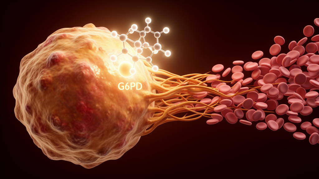
Glucose-6-phosphate dehydrogenase, or G6PD, is best known as the rate-limiting enzyme of the pentose phosphate pathway and as the culprit behind the most widespread enzyme deficiency in the world. A new study published in EMBO Reports on 13 October 2026 now assigns the protein an unexpected second job in blood biology: in both human patients and mice, a lack of G6PD was associated with higher platelet counts, and the researchers traced the effect to a previously unrecognized, non-metabolic role of G6PD in controlling the cytoskeletal choreography that drives platelet production.
The clinical signal came from a retrospective analysis of patients with congenital G6PD deficiency recruited through the Department of Laboratory Medicine of Ruijin Hospital in Shanghai. Among 17 G6PD-deficient male patients aged 18 to 40, average platelet counts were approximately 25 percent higher than in age-matched healthy male controls, and six female patients showed a similar trend. The patients were evaluated in records obtained outside acute hemolytic episodes, and all were free of hematologic disorders and serious underlying disease. The authors also noted an approximately 8 percent reduction in hemoglobin levels and an approximately 6 percent decrease in red blood cell counts in the deficient patients, consistent with the shortened red cell lifespan that characterizes the condition.
To test whether the association reflected a direct effect of G6PD on platelets rather than a secondary consequence of anemia, the team built megakaryocyte- and platelet-specific G6PD knockout mice, in which the gene was deleted using the Pf4-Cre system. These animals carried about 15 percent more platelets in their peripheral blood than floxed controls, while mean platelet volume, red cell counts, and white cell counts were unchanged. Platelet function also appeared intact: aggregation and secretion responses, surface glycoprotein expression, granule content, and platelet ultrastructure were all comparable between knockout and control animals.
Platelet counts reflect a balance between production and clearance, so the researchers dissected both sides of the equation. The proportion of reticulated, newly released platelets was significantly higher in the knockout mice, and after platelets were depleted with an anti-GPIbα antibody, counts recovered significantly faster in the deficient animals. At the same time, the fraction of Annexin V-positive apoptotic platelets and the in vivo lifespan of biotin-labeled platelets were similar in both groups. The conclusion was that G6PD loss raises platelet numbers by accelerating production, not by prolonging platelet survival.
Zooming in on the cellular source of platelets, the team found no difference in plasma thrombopoietin levels or in the number of bone marrow megakaryocytes between the genotypes, and polyploidization of cultured megakaryocytes was unaffected. The action lay further down the differentiation pathway. In a proplatelet formation assay on immobilized fibrinogen, a significantly higher proportion of G6PD-deficient megakaryocytes entered the proplatelet-forming stage, and individual forming cells extended more branches and more proplatelet tips. Electron microscopy revealed a more elaborate demarcation membrane system and more abundant membranous protrusions at the cell periphery in mature deficient megakaryocytes, along with an increased cell size, indicating enhanced cytoplasmic maturation.
Because actin cytoskeletal organization guides proplatelet branching, the investigators stained forming megakaryocytes for F-actin. In control cells, F-actin tended to accumulate in specific regions of the undivided cytoplasm, whereas in G6PD-deficient cells it showed a more uniform spatial distribution, a pattern the authors interpret as facilitated actin remodeling. Deficient platelets spreading on fibrinogen also organized their cytoskeleton more extensively, spread over larger areas, and reached more advanced spreading stages. Notably, within the proplatelet buds themselves, F-actin content and distribution showed no significant differences, localizing the cytoskeletal effect to the megakaryocyte body during proplatelet formation.
Strikingly, the metabolic role of G6PD seemed irrelevant to this phenotype. The ratios of NADPH to NADP+ and NADH to NAD+ were similar in deficient and control platelets, as were the extracellular acidification and oxygen consumption rates measured by Seahorse analysis. Targeted glucose metabolomics detected no significant changes in ribose-5-phosphate or key glycolytic and tricarboxylic acid cycle products in the absence of G6PD. The authors propose that compensatory metabolic routes may sustain key metabolites, and that the platelet phenotype instead stems from a non-canonical, physical function of the enzyme.
That function emerged from co-immunoprecipitation and mass spectrometry of human platelet lysates, which identified the actin-binding protein tropomodulin-3, or TMOD3, as a G6PD-interacting partner; gene ontology analysis of the enriched proteins highlighted actin cytoskeleton organization. TMOD3 caps the pointed ends of actin filaments, stabilizing F-actin while limiting its turnover, and previous work has shown that TMOD3 knockout disrupts F-actin organization in megakaryocytes and impedes proplatelet formation. In the new study, co-immunoprecipitation confirmed the G6PD-TMOD3 interaction in human platelets and HEK 293T cells, and structured illumination microscopy showed the two proteins colocalizing on F-actin bundles in murine megakaryocytes. When G6PD was knocked down in Meg01 cells, the interaction between TMOD3 and actin weakened, actin polymerization increased, and cells spread over larger areas. In G6PD-deficient megakaryocytes, the colocalization of TMOD3 with F-actin, particularly in the F-actin-rich region near the cell periphery, was markedly reduced, with a lower Pearson correlation coefficient between the two signals.
The interaction proved sensitive to NADP+, which serves G6PD both as a coenzyme and as an allosteric regulator, with one G6PD monomer binding one glucose-6-phosphate and two NADP+ molecules. In co-immunoprecipitation assays, glucose-6-phosphate had no effect on G6PD-TMOD3 binding, whereas increasing NADP+ concentrations progressively enhanced the interaction, and NADP+ also increased the colocalization of the two proteins on F-actin bundles at the cellular edge. Functionally, NADP+ reduced the spreading of human platelets, thickened peripheral F-actin bundles in mature control megakaryocytes, and inhibited proplatelet formation in control megakaryocytes while leaving G6PD-deficient cells unaffected, indicating that the inhibitory effect of NADP+ on platelet production runs through G6PD.
Finally, the team mapped the TMOD3-binding sites to the N-terminal NADP+-binding domain and the C-terminal structural NADP+-binding domain of G6PD, screened thirteen derived peptides by ELISA, and found that peptide GC2P2, spanning residues 366 to 395, bound TMOD3 most strongly and strengthened both the G6PD-TMOD3 interaction and TMOD3’s binding to actin. Fused to the HIV-derived cell-penetrating peptide TAT to yield C-GC2P2, the peptide reduced platelet spreading, decreased the proportion of proplatelet-forming megakaryocytes and their branching, and, when injected into mice after platelet depletion, significantly slowed platelet regeneration in control animals around 48 hours after injection, with no effect in G6PD-deficient mice, confirming a G6PD-dependent mechanism. C-GC2P2 did not impair platelet aggregation, granule release, or morphological parameters. The authors suggest that enhancers or inhibitors targeting the G6PD-NADP+-TMOD3 complex could become strategies for thrombocytosis or thrombocytopenia, while noting the study’s limitations: the human data are retrospective and modest in size, and no blinding was performed during statistical analysis, so the findings will need prospective confirmation before clinical translation.
Subject of Research: The role of glucose-6-phosphate dehydrogenase in regulating platelet production through TMOD3-mediated actin dynamics
Article Title: G6PD suppresses thrombopoiesis via interfering with TMOD3-mediated actin dynamics
Article References: Xin, M., Zhang, C., Liang, T., Jin, P., Shao, Y., Chen, C., Li, G., Zhang, K., Jiang, H., Cai, X., Fan, X., Liu, J., Wang, X., Xu, Y., & Dai, J. (2026). G6PD suppresses thrombopoiesis via interfering with TMOD3-mediated actin dynamics. EMBO Reports. https://doi.org/10.1038/s44319-026-00962-7
Image Credits: AI Generated
DOI: 10.1038/s44319-026-00962-7
Keywords: G6PD deficiency, thrombopoiesis, platelets, megakaryocytes, TMOD3, actin dynamics, NADP+, proplatelet formation, pentose phosphate pathway, GC2P2 peptide, platelet biogenesis, thrombocytosis
News Source: Drew Townsend. (October 29, 2026). G6PD Deficiency Raises Platelet Counts by Loosening a Brake on Platelet Production. Scienmag.