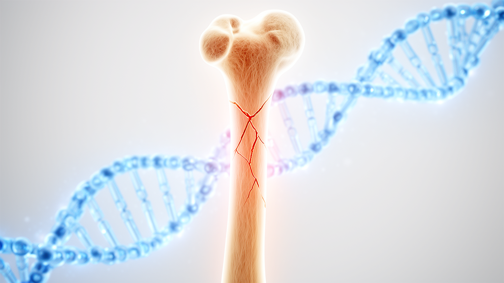
Osteogenesis imperfecta, often called brittle bone disease, is a rare inherited condition that affects roughly one in every 15,000 to 20,000 births. A review published in Frontiers in Cell and Developmental Biology traces how mutations in more than twenty genes disrupt the body's ability to build strong bone and surveys the treatments now in use, from bisphosphonates to experimental cell and gene therapies. The review, written by Giacomina Brunetti of the University of Bari Aldo Moro in Italy, notes that bisphosphonates and monoclonal antibodies represent elective therapy, while trials of cell and gene therapies are ongoing and offer new options for osteogenesis imperfecta patients.
At its core, osteogenesis imperfecta is a disease of type I collagen, the main protein present in the extracellular matrix of bone and the body. Since the early 1980s, the condition was understood as a predominantly inherited disease caused by mutations in either of the two genes that encode type I collagen, COL1A1 and COL1A2. These mutations impair the synthesis and structure of collagen, producing the classic phenotype: low bone mass, bone fragility, and possible fractures. The original Sillence classification recognized four types, ranging from type I, in which patients show near-normal stature, blue sclera, and late-onset hearing loss, to type II, a perinatally lethal form, with type III marked by progressive deformity and type IV by bone deformity of intermediate severity.
What has transformed the field is the discovery that collagen itself is not the whole story. Recent studies have identified mutations in a growing list of collagen-related genes, and the genetic classification now includes 22 types of osteogenesis imperfecta, with a 23rd registered in OMIM in 2023. Each type maps onto a specific step in collagen biology. Types V and VI involve impaired bone mineralization through mutations in IFITM5 and SERPINF1, respectively. Types VII, VIII, and IX disrupt the ER-residing prolyl 3-hydroxylase complex formed by CRTAP, P3H1, and PPIB, which modifies procollagen chains. Types X and XI affect the collagen chaperones HSP47 and FKBP65, while type XII involves BMP1, a metalloprotease that cleaves procollagen into its mature form. A further group, types XIII through XVIII, alters osteoblast activity and differentiation through genes such as SP7, TMEM38B, WNT1, CREB3L1, SPARC, and MBTPS2.
The cellular machinery behind these defects is intricate. Type I procollagen is assembled from two pro-α1 chains and one pro-α2 chain in the endoplasmic reticulum, where it undergoes post-translational modifications at proline and lysine residues. Chaperones like HSP47 and FKBP10 ensure proper folding and trafficking to the Golgi, while the KDEL receptor 2 recycles resident ER proteins. Calcium flux from the ER, regulated by the channel TRIC-B, is essential for these modifications. After secretion, BMP1 and ADAMTS-2 cleave the terminal propeptides, and collagen molecules assemble into fibers stabilized by crosslinks formed through the action of lysyl oxidases. Disruption at any point in this assembly line produces bone that is structurally weaker, and microscopic studies of types II and III show lamellar bone with short, obliquely oriented lamellae and osteoblasts with dilated rough endoplasmic reticulum, signs of cellular stress.
Beyond collagen processing, the review highlights additional mediators that shape the disease. Schnurri-3, a negative modulator of bone formation, has been proposed as a potential target for osteogenesis imperfecta management, since Schnurri-3 knockdown in an oim/oim mouse model of osteogenesis imperfecta rescued bone impairment. Immune cells also contribute: T cells in the oim/oim mouse model display an activated phenotype with elevated secretion of IFNγ and TNFα, and OI mice transplanted with regulatory T cells from wild-type mice displayed an improved bone phenotype due to reduced T cell activation and cytokine production. The RANKL/RANK/OPG signaling axis, which governs osteoclast formation and bone resorption, is dysregulated in patients: increased RANKL and decreased OPG messenger RNA amounts were demonstrated in lymphomonocytes from pediatric patients with OI types I, III, and IV. Osteocytes, the most numerous cells in bone, show increased density in osteogenesis imperfecta, and in oim/oim mice the conditional loss of RANKL in osteocytes resulted in severely increased trabecular bone mass, indicating that osteocytes significantly contribute to the low bone mass observed in OI.
Muscle weakness compounds the skeletal problems. Patients show lower muscle strength and power, greater fatigability, and impaired balance, stemming from reduced muscle mass and poor muscle quality. In the oim/oim mouse model, mitochondrial dysfunction in skeletal muscle correlates with altered whole-body metabolism and skeletal muscle weakness, and myosin heavy-chain fiber-type distribution is abnormal in the soleus muscle, with a reduction in type I myofibers and an increase in type IIa myofibers compared to wildtype littermates. The bone-muscle unit is a critical potential therapeutic target, and in murine models treatment with activin receptor 2B, inhibiting both activin A and myostatin, led to a beneficial effect on the bone of oim/oim, Col1a2+/G610C, and Col1a1+/Jrt mice.
On the therapeutic front, bisphosphonates represent elective treatment, inhibiting bone resorption by osteoclasts and stimulating their apoptosis, with a consequent increase in BMD, decrease in pain, and augmented muscle strength. Neridronate is the only bisphosphonate approved for pediatric use in current management guidelines, while others can be used in adulthood. Monoclonal antibodies have expanded the toolkit: denosumab, which targets RANKL, significantly increased areal bone mineral density in children with osteogenesis imperfecta, and in a year-long study of adults it enhanced aBMD at the lumbar spine and total hip, with efficacy comparable to zoledronic acid but fewer adverse effects. Anti-sclerostin antibodies, including setrusumab, showed a dose-dependent increase in bone formation and resorption with setrusumab treatment in the phase 2b ASTEROID trial in adults with a clinical diagnosis of OI types I, III, or IV, sustaining its phase 3 evaluation. Teriparatide, a parathyroid hormone analog, increased bone mineral density, though a recent trial involving 350 OI patients found that teriparatide plus zoledronic acid did not reduce fracture risk compared with standard care, suggesting the importance of reduced bone quality rather than low bone density in the pathogenesis of fracture.
Anti-TGF-beta therapy represents another frontier. Fresolimumab, in the first-in-human phase I study using a TGFβ antibody, was demonstrated to ameliorate bone mass with increases in lumbar spine aBMD in adult patients with OI type IV. But the most transformative approaches may lie in cell and gene therapy. Two clinical studies administered intravenous histocompatible mesenchymal stromal cells from donors to children with severe osteogenesis imperfecta, and the MSC administration procedures were safe, with no serious adverse events registered. Six months following two MSC infusions, there was an increase in growth velocity, an 80% reduction in the frequency of fractures, and an increase in total bone mineral content in three of the eight children treated. The BOOSTB4 trial is evaluating postnatal or pre- and postnatal administration of allogeneic expanded human first-trimester fetal liver MSCs (BOOST cells) for OI type III or severe type IV, with primary outcome measures focused on safety and tolerability of repeated BOOST cell administration, and further time is required to obtain results.
Gene editing offers the prospect of correcting mutations directly. Researchers have reprogrammed fibroblasts from OI type II patients into induced pluripotent stem cells, and delivery of the correct linear DNA fragment to patient cells resulted in repair of the DNA mutation, with an 84% success rate. Using whole-exome sequencing, investigators identified a novel cis-double-variant c.[175C>T; 187T>A] in COL1A1 from a 5-year-old OI patient, cultured induced pluripotent stem cells from the patient's fibroblasts, and applied CRISPR/Cas9 gene editing to correct the c.187T>A variant. Correcting the c.187T>A variant alone in CRISPR-edited iPSCs was sufficient to alleviate OI phenotypes, as demonstrated by enhanced amounts of COL1A1, COL1A2, and ALP mRNAs and COL1A1 protein. These findings, the review concludes, strongly encourage the use of CRISPR/Cas9 gene editing as a new therapeutic tool, even as routine screening for the disease remains limited to prenatal ultrasonography and genetic testing for families with a known history.
Subject of Research: Genetic mechanisms and emerging therapies for osteogenesis imperfecta, a rare inherited bone fragility disorder
Article Title: Bone in osteogenesis imperfecta: features and therapy
Article References: Brunetti, G. (2026). Bone in osteogenesis imperfecta: features and therapy. Frontiers in Cell and Developmental Biology, 14, Article 1950969. https://doi.org/10.3389/fcell.2026.1950969
Image Credits: AI Generated
DOI: 10.3389/fcell.2026.1950969
Keywords: osteogenesis imperfecta, type I collagen, COL1A1, COL1A2, bisphosphonates, denosumab, setrusumab, CRISPR gene editing, mesenchymal stromal cells, bone fragility, osteoblasts, genetic classification
News Source: Juliet Wilcox. (October 30, 2026). From Brittle Bones to Gene Editing: New Map of Osteogenesis Imperfecta and Its Therapies. Scienmag.