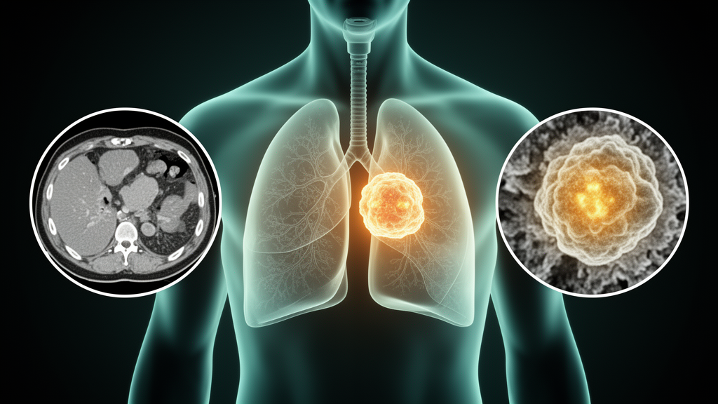
Radiomics promises to turn ordinary medical images into quantitative fingerprints of tumor biology, but the approach has long been haunted by a stubborn problem: the numbers it produces often change every time the scanner, the resolution, or the imaging protocol changes. A new study published in European Radiology Experimental on 7 October 2026 tackles that fragility head-on by asking a deceptively simple question — if you image the same lung tumor on a clinical CT scanner, on an ex vivo CT scanner, and on a microscopic-resolution micro-CT machine, do any radiomic features actually stay the same? The answer, from a retrospective cohort of 60 patients with non-small-cell lung cancer (NSCLC), is a cautious yes, but only for a small handful of texture-based measures.
The research team, led by Leonardo Brizzi and Lorenzo Preda with colleagues across Italian institutions, enrolled 60 NSCLC patients who had undergone surgery without prior therapy. The cohort was 65% male and 35% female, with a mean age of 74.4 ± 7.7 years (range 54–91), and histological subtypes were equally split between adenocarcinoma (ADK) and squamous cell carcinoma (SCC). Each patient contributed three kinds of imaging: a preoperative clinical CT retrieved from the hospital archive, a conventional CT scan of the excised surgical specimen, and an ultra-high-resolution micro-CT scan of the same tissue preserved as a paraffin-embedded block.
The micro-CT workhorse was a SkyScan 1276 scanner operating at 40 kVp and 200 µA, producing isotropic voxels of just 20 × 20 × 20 micrometers — fine enough to resolve structural details invisible to clinical CT, whose in-plane voxel sizes ranged from 0.586 to 1.367 millimeters. The ex vivo CT scans were acquired on a Siemens Somatom Go.Top at 120 kV with 0.6-mm slices. Tumor regions of interest were segmented semi-automatically in ITK-SNAP by an experienced radiologist, with micro-CT contours reviewed in consensus with a pathologist to help define tumor boundaries at the histological scale.
Before any features were computed, the team applied a battery of harmonization steps designed to strip out acquisition-related variability. All three modalities were resampled to standardized isotropic voxels, gray-level distributions of the CT images were normalized to match the median and interquartile range of the corresponding micro-CT regions, and ex vivo CT images were registered to the micro-CT reference using non-rigid B-spline registration. Because paraffin blocks could hold at most about 2 centimeters of tissue while mean tumor diameter was 2.97 ± 1.58 centimeters (range 1.0–7.5), the whole tumor often could not fit into the micro-CT field of view. The researchers therefore repeated their analyses twice: once with whole-tumor segmentations and once with volume-matched subsampled regions carved out of the in vivo CT to mirror the geometry of the excised block.
From each region they extracted 107 radiomic features using Pyradiomics v3.0.1, software compliant with the Image Biomarker Standardisation Initiative. Shape features were excluded because the subsampling destroyed the original tumor geometry, leaving 93 features for analysis. The results were sobering. In non-normalized datasets, the average relative difference between modalities ranged from 399.8% to 596.5%, with first-order intensity features swinging by more than 1,200%. Mean intraclass correlation coefficients (ICC) hovered between 0.01 and 0.12, no feature reached the 0.75 threshold for good agreement, and fewer than 11% of associations reached statistical significance.
Harmonization changed the picture substantially. After gray-level normalization, the variability of first-order features dropped from over 1,200% to roughly 320%, median ICC values rose to approximately 0.20–0.23, and the proportion of features with ICC above 0.5 climbed from under 5% to 12–18%. Mean Spearman correlation coefficients roughly doubled from about 0.10 to about 0.20. Volume matching helped further: subsampled regions raised the significance percentages of features by roughly 8–10 percentage points in both cross-modality comparisons involving in vivo CT. The best-performing configuration was the normalized dataset with subsampled regions, where the ex vivo CT versus subsampled in vivo CT comparison yielded average Spearman and ICC values of 0.23 ± 0.31, with first-order features reaching correlations of 0.51–0.63 while texture features lagged at 0.08–0.14.
Yet even under the most favorable conditions, only a small minority of features could be called acceptably stable. The team’s strict criterion — statistical significance at p ≤ 0.001 across all modality comparisons in both normalized and non-normalized datasets with subsampled regions — identified exactly four survivors, all of them texture measures: DependenceNonUniformity and GrayLevelNonUniformity from the gray-level dependence matrix, GrayLevelNonUniformity from the gray-level run length matrix, and Coarseness from the neighboring gray tone difference matrix. These four features showed Spearman coefficients between 0.579 and 0.792 with p < 0.001, and they retained that stability whether the images came from a hospital CT scanner or a laboratory micro-CT. First-order features were deliberately excluded from this selection because their apparent agreement could simply reflect the normalization procedure itself.
The biological payoff came when the researchers tested whether these stable features could distinguish adenocarcinoma from squamous cell carcinoma. In whole-tumor in vivo CT, all four features separated the two histotypes significantly (p < 0.01), with higher values generally in SCC — except Coarseness, which tended to be higher in ADK. In the volume-matched subsampled setting the same directional trends persisted, though statistical significance weakened to roughly p = 0.05–0.08 for some comparisons and disappeared for others. Across all modalities, including micro-CT, the trend patterns between histotypes remained broadly comparable, supporting the idea that these texture measures capture structural properties of the tumor — cellularity and intratumoral heterogeneity — that survive changes in imaging resolution.
Unsupervised k-means clustering added a further twist. When all 93 features were used, cluster assignments showed no significant association with histotype, whether the data came from in vivo CT (χ² = 0.70, p = 0.402) or micro-CT (χ² = 0.71, p = 0.398). But when clustering was restricted to the four selected stable features, the in vivo CT analysis produced a significant link (χ² = 4.28, p = 0.039), while the micro-CT version did not (χ² = 0.65, p = 0.422). The authors interpret this as evidence that feature stability is not merely a technical nicety but may be a useful filter for building biologically meaningful radiomic models — though they stress that these histotype associations are preliminary and exploratory.
The study’s limitations are candidly laid out. It was a single-center study with a modest sample size and no external validation dataset; micro-CT specimens covered only partial tumor volumes; subsampling may have introduced spatial sampling bias; alignment between in vivo and ex vivo images relied on axial reorientation alone; segmentations were performed by a single operator with no formal inter-observer variability assessment; in vivo CT protocols were heterogeneous; and, crucially, there was no direct quantitative histopathological validation against measures such as cellularity, stromal fraction, or necrosis. Overall reproducibility across acquisition settings remained limited, and the authors conclude that intensity normalization and volume matching are essential prerequisites for any cross-modality radiomic analysis. Their four stable texture features are offered not as validated biomarkers but as candidates for further investigation — a bridge, still under construction, between the radiologist’s image and the pathologist’s slide.
Subject of Research: Cross-modality reproducibility of radiomic features between clinical CT and micro-CT in non-small-cell lung cancer
Article Title: Bridging radiomics and histopathology: cross-modality consistency of radiomic features from CT to micro-CT in NSCLC
Article References: Brizzi, L., Bortolotto, C., Cabini, R. F., Brero, F., Megalizzi, S., D’Ambrosio, G., Filibian, M., Gioia, R., Falvo, P., Stella, G. M., Bottinelli, O. M., Lascialfari, A., & Preda, L. (2026). Bridging radiomics and histopathology: cross-modality consistency of radiomic features from CT to micro-CT in NSCLC. European Radiology Experimental, 10(1), Article 150. https://doi.org/10.1186/s41747-026-00780-5
Image Credits: AI Generated
DOI: 10.1186/s41747-026-00780-5
Keywords: radiomics, micro-CT, NSCLC, lung cancer, texture features, image harmonization, histopathology, reproducibility, adenocarcinoma, squamous cell carcinoma, tumor heterogeneity, biomarkers
News Source: Nathaniel Bowman. (October 29, 2026). Four Radiomic Texture Features Survive the Leap from Clinical CT to Micro-CT in Lung Cancer. Scienmag.