Surgeons at Weifang Eye Hospital in China have reported that three patients with unusually difficult retinal detachments regained retinal reattachment after a treatment built around a foldable capsular buckle, a soft silicone device that indents the eye from the outside. The report, published in Frontiers in Medicine, describes a 71-year-old man with a total detachment involving a small tear and proliferative vitreoretinopathy, a 47-year-old man with a large horseshoe tear exceeding 60 degrees that involved the macula, and a 41-year-old man with a giant retinal tear of roughly 30 disc diameters. All three were treated with the foldable capsular buckle combined with gas tamponade and laser photocoagulation, and the third patient also received minimally invasive vitrectomy. The authors stress that the results reflect the combined strategy rather than the buckle alone, and that the small, uncontrolled nature of the series means no conclusions about effectiveness or superiority can be drawn.
Retinal detachment is a serious, vision-threatening condition in which the neurosensory retina separates from the underlying retinal pigment epithelium. It arises when retinal tears, vitreoretinal traction and liquefied vitreous allow fluid to pass through breaks into the space beneath the retina. Rhegmatogenous retinal detachment, the most prevalent form, is classified as simple or complex according to the location and extent of the detachment and the presence of complications such as proliferative vitreoretinopathy, a condition in which proliferative membranes on the retinal surface generate traction that can frustrate surgical repair. In this report, the authors defined complex detachments as those featuring giant retinal tears, breaks too large for conventional rigid explants to support, total or macula-involving detachment, or grade C proliferative vitreoretinopathy.
Conventional scleral buckling remains the preferred treatment for uncomplicated rhegmatogenous detachment, and it is generally considered ideal for young patients with phakic eyes and limited detachment, particularly when lattice degeneration or inferiorly located tears are present. However, conventional buckling often fails to provide sufficient and precise support for giant or posteriorly located tears. For more complex cases, pars plana vitrectomy has become the standard approach, but it carries drawbacks: accelerated cataract progression in phakic eyes, increased risk of proliferative vitreoretinopathy and epiretinal membrane formation, altered oxygen distribution within the vitreous cavity, and high economic costs. Vitrectomy with silicone oil tamponade also obligates a second operation for oil removal, and the procedure typically requires prolonged postoperative positioning.
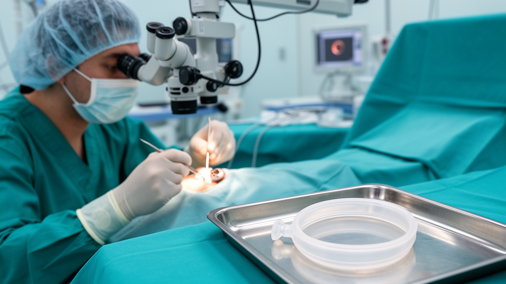
The foldable capsular buckle is a commercially available device modified from the foldable capsular vitreous body AV-10P model manufactured by Vesber in Guangzhou, China. Made of silicone rubber, it consists of a thin, vitreous chamber-shaped capsule with a tube-valve system, weighs approximately 0.3 grams, and shows favorable biocompatibility. Sterile balanced salt solution injected through the valve inflates the capsule, which is implanted on the scleral surface to achieve indentation. Fixation requires only one temporary, superficial suture, in contrast to the multiple intrascleral suture passes needed to anchor conventional explants, making the procedure less traumatic and technically simpler than traditional buckling, with less intraoperative and postoperative discomfort. The valve system also permits intraoperative titration of the indentation force.
Preoperative planning in the series included three-dimensional reconstruction of the detachment configuration to determine the optimal implantation position and inflation volume. Selection criteria required a primary retinal break amenable to focal buckling support, no additional breaks in other quadrants requiring extensive circumferential treatment, and proliferative vitreoretinopathy of grade C or below, with any grade C proliferation localized near the primary break and judged amenable to counteraction by buckle support combined with gas tamponade and, when necessary, limited vitrectomy. All three patients were informed of the risks and benefits of both pars plana vitrectomy with silicone oil and the foldable capsular buckle approach, and all expressed a strong preference for avoiding silicone oil tamponade, prolonged prone positioning and secondary removal surgery. The decision to proceed was made through shared decision-making.
The surgeries were performed by the same experienced clinician under retrobulbar and supraorbital anesthesia. The surgeon incised the bulbar conjunctiva 5 millimeters behind the limbus, created a narrow episcleral tunnel approximately 12 to 14 millimeters long, and released subretinal fluid through a 25-gauge needle. After localizing the retinal break with scleral indentation under an operating microscope, the surgeon placed preplaced sutures of 5-0 nonabsorbable material 4 millimeters anterior to the posterior margin of the break. The foldable capsular buckle was tested for leakage with sterile air, folded, and inserted through the tunnel with its spherical surface facing the sclera. Approximately 1 milliliter of balanced salt solution was injected through the tube valve, which was secured with a nonabsorbable suture, and the position of the break was reconfirmed at the apex of the buckle.
The clinical outcomes were favorable in all three cases. Case 1, the 71-year-old man, presented with blurred vision, a 1 disc diameter round tear at the 1:30 peripheral position, and grayish retinal elevation throughout 360 degrees accompanied by grade C proliferative membranes. He underwent foldable capsular buckle surgery with subretinal fluid release and intravitreal gas injection on December 29, 2024, followed by laser photocoagulation the next day. By the third postoperative day his best corrected visual acuity had improved to 0.3, and at one year the retina remained well adhered with the buckle retained in situ. The authors note that this eye fell outside the grade-based indications used in previous foldable capsular buckle studies, but that the proliferation was localized to the vicinity of the primary break and could be counteracted by the combined strategy.
Case 2, the 47-year-old man, had a 20 disc diameter horseshoe tear in the temporal quadrant with macula-involving detachment in a highly myopic eye with a refractive error of minus 9.00 diopter sphere. High myopia has traditionally been regarded as a relative contraindication to scleral buckling because of long axial length, thin sclera and posteriorly located tears, and the thin sclera increases the risk of perforation during deep scleral suturing for conventional buckles. The foldable capsular buckle requires only an episcleral tunnel and a single superficial suture, minimizing this risk. He underwent surgery on June 18, 2025, with postoperative laser on days 1 and 5. The buckle was removed on postoperative day 50, and six months after removal the retina remained attached with no recurrent detachment, with best corrected visual acuity of 0.6.
Case 3, the 41-year-old man, presented with a giant tear of approximately 30 disc diameters at the 12 o’clock position with an inverted posterior flap and grade C proliferative vitreoretinopathy. The inverted flap generates persistent traction vectors that resist spontaneous reattachment and predispose to redetachment. Minimally invasive vitrectomy was therefore performed to release the traction and relieve the inverted flap, while the foldable capsular buckle provided a broad, spherical compression ridge to mechanically oppose the elastic recoil of the flap and maintain the tear edge in apposition to the retinal pigment epithelium. Early postoperative optical coherence tomography showed persistent subretinal fluid at the tear margin, which the authors attribute to chronic tissue stiffness and slow absorption of viscous fluid rather than surgical failure; they note that in a prospective series, such residual fluid persisted in over 80 percent of eyes at one month after anatomically successful buckling. The buckle was removed on postoperative day 40, and five months later the retina remained attached with no recurrent detachment.
The authors emphasize that the foldable capsular buckle was not evaluated as a standalone intervention in any of the three cases, since all patients received concomitant gas tamponade and laser photocoagulation, and Case 3 underwent additional vitrectomy. The observed anatomical outcomes therefore cannot be attributed to the buckle in isolation. No serious intraoperative or postoperative complications were observed, and the only procedure-related adverse effect was an anticipated refractive shift, which partially resolved in Cases 2 and 3 and persisted with the buckle retained in Case 1. Potential risks documented in previous studies include transient intraocular pressure elevation, balloon displacement or leakage, conjunctival erosion over the tube valve, transient diplopia, and scleral perforation during suture placement in eyes with thinned sclera. The absence of complications in this small series likely reflects careful patient selection, and larger cohorts are needed to fully establish the safety profile. The authors conclude that the approach may be feasible for appropriately selected patients who wish to avoid silicone oil tamponade, but that the appropriate boundary between this strategy and pars plana vitrectomy with silicone oil remains to be defined prospectively.
Subject of Research: A case series evaluating foldable capsular buckle scleral buckling combined with gas tamponade and laser photocoagulation for complex retinal detachment
Article Title: Case Report: Foldable capsular buckle scleral buckling for complex retinal detachment: a case series of three patients
Article References: Liu, X., Wang, W., & Zhang, J. (2026). Case Report: Foldable capsular buckle scleral buckling for complex retinal detachment: a case series of three patients. Frontiers in Medicine, 13, Article 1945579. https://doi.org/10.3389/fmed.2026.1945579
Image Credits: AI Generated
DOI: 10.3389/fmed.2026.1945579
Keywords: retinal detachment, foldable capsular buckle, scleral buckling, pars plana vitrectomy, proliferative vitreoretinopathy, giant retinal tear, gas tamponade, laser photocoagulation, rhegmatogenous retinal detachment, ophthalmology, case series, silicone oil tamponade
News Source: Ophelia Keating. (October 28, 2026). Foldable Capsular Buckle Shows Promise in Three Complex Retinal Detachment Cases. Scienmag.