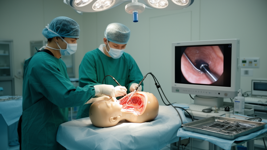
Neurosurgeons training to operate inside the brain’s fluid-filled ventricles have long faced a stubborn problem: once cerebrospinal fluid drains from a body donor, the ventricles collapse, and with them goes the realistic anatomy that makes simulation worthwhile. A team of researchers reports in Acta Neurochirurgica a new way around that obstacle, using a gravity-driven filling system to keep donor ventricles continuously distended throughout endoscopic procedures.
The study, led by Francesco Tengattini and colleagues across European institutions, set out to create a continuous fluid-filled body donor model that recreates a realistic intraventricular environment for anatomical research, neuroendoscopic simulation, and surgical training. The work is published as an anatomic demonstration, meaning it establishes and validates the model itself rather than reporting outcomes in patients.
Five fresh body donor heads were prepared using a gravity-driven ventricular filling system connected through a frontal external ventricular catheter. This arrangement allowed fluid to be infused continuously, counteracting the collapse that normally follows cerebrospinal fluid loss and preserving the physiological shape of the ventricular system during the entire procedure.
Through a mini-corticectomy, the team performed a fully endoscopic transcortical approach using exclusive endoscopic visualization and bimanual microsurgical dissection. In practical terms, both of the surgeon’s hands worked through a single surgical corridor while a camera provided the only view, with no static retractors holding tissue aside. Anatomical exposure, surgical manoeuvrability, and overall feasibility of the model were assessed qualitatively.
The results support the central premise of the design. Continuous gravity-driven infusion consistently restored and maintained ventricular distension, preserving ventricular anatomy throughout the procedures. Successful ventricular cannulation was achieved in all five specimens, although procedural completion was reached in three of the five heads, a detail the authors report transparently.
Within those distended ventricles, the endoscope delivered high-quality visualization of the lateral and third ventricles, enabling detailed identification of the principal intraventricular landmarks that surgeons must recognize during real operations. That imaging quality matters for training, because anatomical fidelity is precisely what is lost when fluid drains and structures deform in conventional cadaveric setups.
The bimanual technique tested within the model also performed as intended. The approach allowed unrestricted bimanual instrument manipulation through a single surgical corridor under continuous endoscopic visualization, without the need for static retractors. The authors conclude that, within this environment, the minimally invasive endoscopic approach proved feasible and reproducible while preserving that freedom of two-handed microsurgical work.
In their conclusions, the researchers position the model as a realistic and reproducible platform for neuroendoscopic anatomical research, surgical simulation, and training. Beyond preserving ventricular anatomy, the setup enables high-quality endoscopic documentation and facilitates the evaluation of novel intraventricular techniques before they are considered in clinical settings.
Several boundaries of the work deserve emphasis. The findings come from five cadaveric heads, with qualitative assessment of exposure and manoeuvrability rather than quantitative performance measures, and the model was used to demonstrate feasibility of a new technique, not to establish its safety or efficacy in patients. The article is also being shared early as a peer-reviewed accepted manuscript, subject to further editorial processing before the final version of record.
Even with those limits, the contribution addresses a recognized gap. As the authors note, realistic body donor anatomical simulation of intraventricular neuroendoscopy has remained limited precisely because ventricular collapse prevents preservation of physiological anatomy. By keeping fluid flowing, the new model keeps the anatomy alive in the only sense a donor specimen allows, offering trainees and researchers a more faithful rehearsal space for some of the most delicate corridors in neurosurgery.
Subject of Research: A fluid-filled cadaveric model for full endoscopic intraventricular surgical simulation and training
Article Title: Development of a novel continuous fluid-filled cadaveric model for full endoscopic intraventricular surgical simulation: anatomic demonstration
Article References: Tengattini, F., De Deus da Rocha Ribeiro Gonçalves, V., Agosti, E., De Notaris, M., Panciani, P. P., Fiorindi, A., Fontanella, M. M., Prats-Galino, A., Di Somma, A., & Enseñat, J. (2026). Development of a novel continuous fluid-filled cadaveric model for full endoscopic intraventricular surgical simulation: anatomic demonstration. Acta Neurochirurgica. https://doi.org/10.1007/s00701-026-07069-x
Image Credits: AI Generated
DOI: 10.1007/s00701-026-07069-x
Keywords: neuroendoscopy, surgical simulation, cadaveric model, ventricular system, cerebrospinal fluid, surgical training, anatomy, minimally invasive surgery, body donor, third ventricle, bimanual dissection, neurosurgery
News Source: Ophelia Keating. (October 28, 2026). Fluid-Filled Cadaver Model Keeps Ventricles Realistic for Endoscopic Surgery Training. Scienmag.