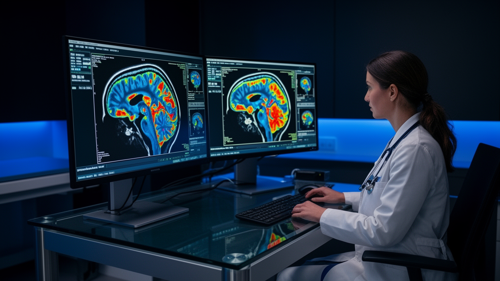
In a histopathology-validated study evaluating 472 tissue samples from 61 patients with IDH-wildtype glioblastoma, O-(2-[¹⁸F]fluoroethyl)-L-tyrosine positron emission tomography ([18F]FET PET) depicted tumor tissue significantly better than contrast-enhanced magnetic resonance imaging (CE-MRI), particularly within regions showing no contrast enhancement.
Accurate visualization of glioblastoma remains essential for guiding surgical resection, biopsy, and radiotherapy planning. While contrast-enhanced MRI is standard in routine clinical practice, it can fail to detect metabolically active tumor tissue situated outside areas of blood-brain barrier disruption.
In diagnostic performance assessments across the analyzed tissue specimens, [18F]FET PET achieved a sensitivity of 91.2% for tumor detection compared to 52.6% for contrast-enhanced MRI. Furthermore, [18F]FET PET maintained high diagnostic reliability in non-enhancing brain tissue, demonstrating a positive predictive value of 98.3%.
These diagnostic findings proved consistent across both newly diagnosed and recurrent disease settings, as well as in patient cohorts undergoing either surgical resection or stereotactic biopsy. According to the study authors, [18F]FET PET complements conventional MRI by uncovering MRI-occult tumor burden, indicating that integrating amino acid PET into routine clinical workflows may improve tumor visualization accuracy and patient management.
The investigation addressed a recognized challenge in neuro-oncology: contrast-enhanced magnetic resonance imaging is routine in clinical practice, yet it can miss metabolically active tumor outside areas of blood-brain barrier disruption. To evaluate whether amino acid imaging could overcome this limitation, researchers assessed O-(2-[¹⁸F]fluoroethyl)-L-tyrosine positron emission tomography ([18F]FET PET) in a histopathology-validated cohort involving 472 tissue samples from 61 patients with IDH-wildtype glioblastoma. Direct comparison with contrast-enhanced MRI showed that [18F]FET PET depicts tumor tissue significantly better than contrast-enhanced scans, especially within regions that display no enhancement on MRI.
The quantitative diagnostic metrics highlighted a substantial difference in detection performance between the two modalities. Across the examined tissue specimens, [18F]FET PET achieved a sensitivity of 91.2% for tumor detection, compared to 52.6% for contrast-enhanced MRI. In addition to this substantially higher sensitivity, [18F]FET PET maintained high reliability in non-enhancing tissue, yielding a positive predictive value of 98.3%. These findings indicate that positive amino acid PET signals consistently identify tumor tissue even where standard contrast agents fail to accumulate.
Crucially, the diagnostic consistency of [18F]FET PET persisted across different patient subsets and clinical stages. The results remained consistent between patients with newly diagnosed glioblastoma and those presenting with recurrent disease. Furthermore, comparable diagnostic performance was documented across both patient cohorts undergoing surgical resection and those undergoing biopsy. This broad applicability suggests that the imaging modality provides dependable information regardless of whether a patient presents at initial diagnosis or with recurrence, and irrespective of the specific neurosurgical procedure performed.
Because accurate visualization of glioblastoma is essential for guiding biopsy, resection, and radiotherapy, uncovering MRI-occult tumor burden carries important clinical implications. By demonstrating that [18F]FET PET complements magnetic resonance imaging through the detection of previously unseen tumor tissue, the study supports incorporating amino acid PET into routine treatment planning. Adopting this imaging approach may ultimately improve the precision of tumor delineation and optimize subsequent patient management strategies.
Subject of Research: Diagnostic accuracy of FET PET in detecting non-contrast-enhancing glioblastoma
Article Title: [18F]FET PET enables histopathology-validated detection of non-contrast-enhancing glioblastoma
Article References: Stürzl, R., Kirchleitner, S. V., Lorenz, J., Schmidbauer, K. J., Stöcklein, V., Quach, S., Kaiser, L., Stöcklein, S., Zounek, A. J., Barci, E., von Polenz, I., Blobner, J., Greve, T., Brendel, M., Harter, P., Ruf, V. C., Rupprecht, R., Ringel, F., Kalasauskas, D., … Albert, N. L. (2026). [18F]FET PET enables histopathology-validated detection of non-contrast-enhancing glioblastoma. Nature Communications, 17(1), Article 11498. https://doi.org/10.1038/s41467-026-78980-1
Image Credits: AI Generated
DOI: 10.1038/s41467-026-78980-1
Keywords: glioblastoma, [18F]FET PET, contrast-enhanced MRI, neuro-oncology, molecular imaging, IDH-wildtype glioblastoma, histopathology, brain tumor imaging, enables, histopathology-validated, detection, non-contrast-enhancing
News Source: Nathaniel Bowman. (November 10, 2026). FET PET Detects Non-Enhancing Glioblastoma In Tissue-Validated Study. Scienmag.