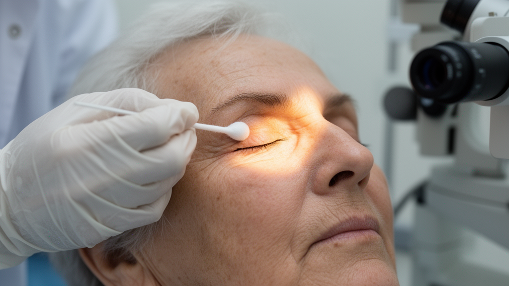
Long-term users of topical eye medications show distinct patterns of periocular change, and the type of medication appears to matter, according to a cross-sectional study published in BMC Ophthalmology. Researchers at Göztepe Prof. Dr. Suleyman Yalcin City Hospital in Istanbul, Türkiye, examined 115 patients who had been using topical ophthalmic treatments continuously for at least one year, along with 50 healthy controls aged 45 to 75 years. Preservative-containing treatment, by contrast, showed an exploratory association with lower eyelid herniation. Notably, the study found no clear associations between eyelid manipulation during eye-drop instillation and any of the evaluated periocular outcomes after correction for multiple comparisons.
The treatment groups reflected routine clinical populations rather than study-assigned interventions: 50 participants used only antiglaucoma drops, 35 used only artificial tears for dry eye disease, and 30 used both. Participants in the antiglaucoma groups had confirmed primary open-angle glaucoma, verified through clinical records, optic nerve head evaluation, retinal nerve fiber layer optical coherence tomography, standard automated perimetry, and gonioscopy. The artificial tear groups had documented dry eye symptoms confirmed by tear break-up time and Schirmer testing. Healthy controls had no diagnosis, symptoms, or clinical evidence of either condition and no history of regular topical eye-drop use.
After adjustment for age and sex, all three treatment groups showed lower margin reflex distance 1 values than controls, with all comparisons remaining significant after false discovery rate correction, and all treatment groups showed higher frequency and severity of upper eyelid atrophy than controls. The antiglaucoma group additionally had lower levator function, with an adjusted mean difference of minus 1.09 millimeters compared with controls. Importantly, upper eyelid changes appeared even in the artificial tear group, which had no prostaglandin analog exposure, suggesting that the upper eyelid findings cannot be attributed solely to prostaglandin use. The extent to which dry eye disease itself, chronic ocular surface inflammation, or other factors contributed to these structural changes remains uncertain, the authors noted.
The pharmacological exposure analysis, restricted to the 80 glaucoma-treated participants, produced the study’s most distinctive pattern. Current prostaglandin analog use was associated with roughly four-fold higher odds of upper eyelid atrophy, with an odds ratio of 4.30 and a 95 percent confidence interval of 1.52 to 12.13, and with more advanced atrophy stages, with a common odds ratio of 5.89. At the same time, prostaglandin analog users had markedly lower odds of visible upper eyelid herniation, odds ratio 0.21, and lower eyelid herniation, odds ratio 0.13. For lower eyelid herniation stage, threshold-specific analyses showed that the inverse association was confined to the presence-versus-absence threshold, with no clear association for progression to more advanced stages. The authors observed that this combination of greater upper eyelid atrophy and reduced visible fat herniation is consistent with the periorbital fat-volume loss phenotype characteristic of prostaglandin-associated periorbitopathy, although the study does not establish the underlying mechanism.
Preservative exposure told a different story. Among eye-drop users, the presence of at least one preservative-containing medication was associated with higher odds of lower eyelid herniation, with an odds ratio of 5.46 and a 95 percent confidence interval of 1.76 to 16.94, and with more advanced lower eyelid herniation stage, with a common odds ratio of 5.10. The number of preservative-containing medications showed similar associations. These estimates carried relatively wide confidence intervals, indicating limited precision, and the authors emphasized that they should be considered exploratory because preservative type, concentration, and cumulative preservative exposure were not recorded. Crucially, the preservative associations remained significant after mutual adjustment for prostaglandin analog exposure, and the prostaglandin findings remained significant after mutual adjustment for preservative exposure, supporting the internal consistency of the two exposure-phenotype patterns within the limitations of the cross-sectional design.
Perhaps the most striking null result concerned eyelid manipulation. Patients frequently retract the upper or lower eyelid to facilitate drop administration, and repeated mechanical manipulation has been hypothesized to contribute to periocular structural changes, with prior evidence extrapolated from contact lens wear and chronic eyelid taping. After false discovery rate correction, no clear associations emerged between either the cumulative exposure measure or the manipulation patterns and any of the evaluated periocular outcomes. A nominal inverse association between cumulative exposure and levator function did not survive correction.
The authors were careful about what this null result does and does not mean. They stressed that the findings should not be interpreted as evidence that mechanical factors play no role in periocular changes. Rather, the estimated manipulation-weighted exposure measure, which was derived from self-reported behavior, daily drop frequency, and treatment duration, did not capture the magnitude or direction of the applied force, unsuccessful instillation attempts, contact duration, or changes in technique over time. The analyses also assumed that the instillation technique reported at the single study visit had remained stable throughout the treatment period. Further prospective studies using more direct measures of mechanical exposure are warranted, the authors concluded.
Several further limitations frame the results. Because periocular findings were assessed at a single time point after long-term treatment, the onset and temporal progression of the structural changes could not be determined, and no causal inferences can be drawn. The examining ophthalmologist was not masked to participants’ diagnoses or treatments, and intraobserver and interobserver agreement for the photographic assessments were not formally evaluated. Because the groups were defined by underlying clinical conditions, the effects of the medications could not be completely separated from those of glaucoma or dry eye disease. Previous prostaglandin analog use and the duration of the current prostaglandin regimen were not recorded, so the pharmacological analyses reflect current medication exposure at enrollment rather than cumulative medication-class exposure. Relatively small sample sizes in some medication subgroups may have limited the ability to detect weaker associations.
Within those constraints, the study offers a consolidated picture that previous work, which generally examined pharmacological, preservative, and mechanical factors separately, had lacked. The authors concluded that different treatment-related exposures may be associated with different aspects of periocular morphology: prostaglandin analog exposure with a fat-volume loss phenotype in the upper eyelid and reduced visible herniation, preservative exposure with lower eyelid herniation, and neither with malar festoons, for which no clear associations were identified after correction. They called for prospective longitudinal studies incorporating standardized exposure assessment, detailed preservative characterization, and histopathological investigations to confirm the findings and clarify the biological mechanisms. Until such studies are available, the reported associations should be read as cross-sectional exposure-phenotype patterns in long-term topical treatment users, not as demonstrated causal effects.
Subject of Research: Associations of prostaglandin analogs, preservatives, and eyelid manipulation with periocular morphology in long-term users of topical ophthalmic medications
Article Title: Periocular morphology during long-term topical ophthalmic treatment: associations with prostaglandin analogs, preservatives, and eyelid manipulation
Article References: Karaca, S., Yilmaz, O. F., Sahin, Z. B., Kaya, S. P., Kirik, F., Hepokur, M., & Oguz, H. (2026). Periocular morphology during long-term topical ophthalmic treatment: associations with prostaglandin analogs, preservatives, and eyelid manipulation. BMC Ophthalmology, 26(1), Article 692. https://doi.org/10.1186/s12886-026-05485-2
Image Credits: AI Generated
DOI: 10.1186/s12886-026-05485-2
Keywords: glaucoma, prostaglandin analogs, periocular morphology, upper eyelid atrophy, eyelid herniation, preservatives, dry eye disease, eye drops, levator function, prostaglandin-associated periorbitopathy, cross-sectional study, BMC Ophthalmology
News Source: Ophelia Keating. (October 28, 2026). Eye Drops and Eyelids: Prostaglandins Tied to Upper Lid Atrophy in Cross-Sectional Study. Scienmag.