Credit: Chulhong Kim (POSTECH)
Photoacoustic imaging has gained global attention for capturing images without causing pains or using ionizing radiation. Recently, many researchers have heavily studied on observing deep tissues to apply the photoacoustic imaging to clinical diagnosis and practices.
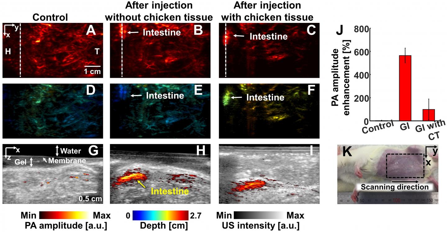
Prof. Chulhong Kim of Creative IT Engineering from POSTECH and his student, Byullee Park conducted joint research with Prof. Hyungwoo Kim and Kyung Min Lee of Cheonnam National University and proposed a new contrast agent for the photoacoustic imaging of deep tissues. They used a nickel-based nanoparticle as a contrast agent that absorbs light at 1,064 nm wavelength. They obtained images of the tissues penetrated in maximum 3.4 cm depth in live animals which is the deepest image observed using this wavelength compared to the previous studies.
The principle of photoacoustic imaging is that it allows lights to be absorbed by tissues which then causes a thermoelastic expansion of the tissues of the organs instantly. This generates sound wave (photoacoustic) signals that are detected as ultrasound wave sensors, producing images. The conventional optical microscopic imaging technologies only allow observing tissues in 1 mm depth. On the other hand, the photoacoustic imaging system produces images of the deep tissues in animals and humans based on optical contrast.
However, the photoacoustic imaging is challenging despite of intense research activities to observe deep tissues in various organs more closely. It is difficult to deliver enough light at 650~900nm short-wavelength with an affordable cost to deep tissues in the body. For this reason, the commercial and clinical translation of photoacoustic imaging are challenging.
To improve this limitation of the photoacoustic imaging, the research team introduced a nanoparticle nickel-based contrast agent, that absorbs light specifically and strongly at 1,064 nm wavelength, to observe deep tissues. They verified biocompatibility of nickel-based nanoparticles and obtained photoacoustic images in deep tissues (3.4cm depth) of lymph nodes, gastrointestinal tracts, bladders of live rats by inserting the nanoparticles.
The first author of the paper, Byullee Park said, “This research is different from the previous studies that used short wavelength. We used long wavelength lasers and were able to minimize damages in the tissues. We were also able to obtain images of deep tissues by delivering lights to organs located in deep inside of the animal.”
When this newly developed photoacoustic imaging technique is applied to clinical practices, it can help diagnosis of diseases related to deep organs by producing images noninvasively and without a risk of explosion to radiation unlike other imaging methods that need radiations such as computed tomography (CT). Furthermore, lasers of 1,064 nm wavelength are comparatively economical, and can be used with other commercial ultrasound machines, which bring anticipation of its early clinical applications.
“Our research is the first example of imaging the deepest tissues in the body among all the research papers on photoacoustic imaging so far. It is very meaningful that it has taken a step further to clinical feasibility of photoacoustic imaging,” said Prof. Chulhong Kim, the corresponding author of the paper.
###
This research was financially supported by ICT Consilience Creative Program of the Ministry of Science and ICT, the Korea Health Technology R&D Project funded by the Ministry of Health, and Welfare and the Pioneer Project sponsored by the National Research Foundation. Details and conclusions of this research was introduced as the cover story in the international journal of nanoparticle imaging diagnosis and treatment, Theranostics.
Media Contact
Jinyoung Huh
[email protected]
82-542-792-415
Original Source
http://postech.
Related Journal Article
http://dx.


