A groundbreaking development in bioimaging has emerged from the innovative research team at Toyohashi University of Technology, under the guidance of Professor Kazuaki Sawada and Project Assistant Professor Hideo Doi. They have unveiled an advanced semiconductor sensor that opens new avenues for real-time observation of biomolecules in solution, specifically focusing on the dynamics of hydrogen ions and lactate, which are crucial for various biological processes. This cutting-edge technology utilizes semiconductor processes to create a highly efficient imaging system capable of providing invaluable insights into the intricate behaviors of these biomolecules, which are fundamental to understanding neurochemical signaling and brain function.
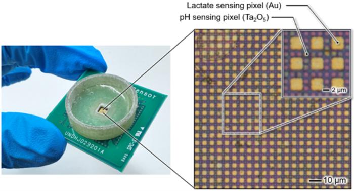
The sensor is uniquely designed with a thin metal film that acts as a neurotransmitter-sensitive membrane, delicately patterned on sensor pixels spaced at just a 2 μm pitch. By employing state-of-the-art semiconductor technology, the researchers have enabled real-time capture of ionic movement as image data. This remarkable advancement boasts a time resolution in the milliseconds range and a spatial resolution of several microns – a scale that is impressively around 1/17 the width of a human hair. Such precision renders the sensor an invaluable tool for measuring the dynamic relationship between neurotransmitter distribution and ionic fluctuations, providing a window into biochemical interactions that occur between neurons in the brain.
Historically, the methods available for detecting these chemical signals have been limited. Conventional devices typically rely on single-point electrodes, which offer limited spatial resolution for measuring biomolecule dynamics. These electrodes, often several tens of microns in diameter, are insufficient for the high-resolution demands of modern neurobiology. Moreover, configurations that utilize arrays of electrodes spaced hundreds of microns apart severely restrict researchers’ abilities to glean detailed spatial and temporal insights into neurochemical phenomena. This novel semiconductor bioimage sensor overcomes these limitations, as it facilitates real-time, simultaneous measurement of multiple biomolecule types with unprecedented spatial accuracy.
Professor Sawada’s laboratory previously achieved similar but less comprehensive goals by creating a semiconductor image sensor to capture ionic movements akin to a camera. The evolution from this earlier work to the current imaging device exemplifies a significant leap in capability. The researchers have managed to create a sophisticated multisensing device where metal electrodes form a lattice-like structure on the sensor array, spaced at 6 μm intervals. This novel configuration allows for real-time simultaneous measurements of both lactate and hydrogen ions, essential for understanding memory formation processes. The incorporation of selective recognition elements, such as enzymes that detect specific biomolecules, enables the measurement of diverse types of neurotransmitters simultaneously.
This expansion from single-point measurements to capturing multiple types of biomolecules signifies a historic achievement in neurobiological research. The ability to visualize and measure two distinct chemical entities with high spatial resolution enables scientists to explore previously unobservable chemical signaling and interactions that occur in intricate biological contexts. It equips researchers with the capability to examine chemical signals in microscopic regions, including the critical interface between neurons, greatly enhancing our understanding of neurotransmission.
A recent collaborative study involving experts from the Faculty of Medicine at the University of Yamanashi further highlights the sensor’s capabilities. The research focused on understanding how hippocampal cells—a key player in memory formation—respond to pharmacological stimuli. The team successfully employed the sensor to concurrently measure lactate release and changes in the extracellular pH. This ability to visualize real-time biochemical dynamics directly within living tissues without any form of labeling represents a paradigm shift in how scientists can approach the study of live cellular processes and interactions.
Moreover, this innovative ion image sensor technology could pave the way for various applications, ranging from basic neuroscience to developing diagnostics for neurological disorders. By providing the tools
Tags: advanced semiconductor sensorsbiomolecule observation techniquesbrain function analysis toolshigh-resolution biochemical measurementshydrogen ions and lactate dynamicsinnovative imaging systems for biologyionic movement imagingneurochemical signaling researchneurotransmitter-sensitive membrane designreal-time bioimagingsemiconductor imaging technologyToyohashi University of Technology research